

Three cases of lesions often seen in primary care, 2 seen in uncommon locations. Test your visual diagnostic skills.
Three cases of lesions often seen in primary care, 2 seen in uncommon locations. Test your visual diagnostic skills.
An up-close look and a short lesson on each of 7 dermatologic lumps and bumps. Would any of them give you cause for alarm?
6 patients who presented with lumps, bumps, and spots. Your diagnoses?

Can you identify these 4 lesions seen in a primary care setting? Should biopsy be performed on any? All? Test your visual diagnostic skills.

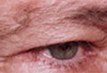
Three men over age 50 years, three different lesions: which would concern you?
In this diagnostic quiz: 3 photos of classic presentations of 3 different diseases in patients of 3 generations.
No labs, no biopsies, no imaging -- only your seasoned clinical judgement is required to make a diagnosis in this case.
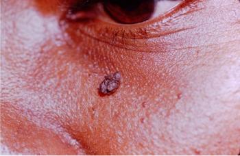
Get an upfront view of irritating dermatologic conditions that often occur on and around the face.
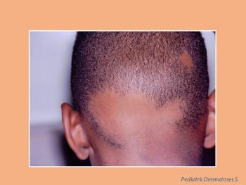
What's the matter with these kids? Get a close look at and a brief overview of seven common dermatoses seen in childhood.
Would any of the 7 lumps and bumps here give you cause for alarm? Get an up-close look and a short lesson on each.
This lesion on the posterior neck of a young woman was present since birth and recurred after electrodessication. Can you Dx?
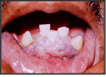
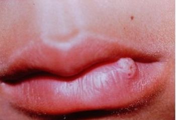
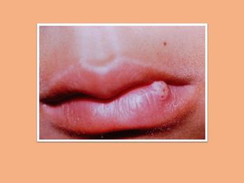
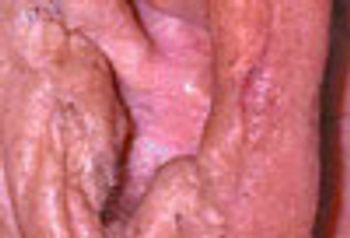
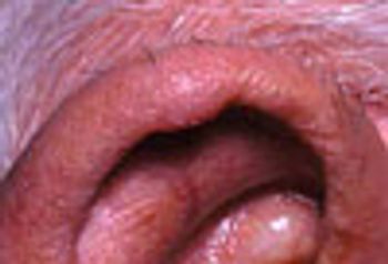
What is in your differential diagnosis for this lesion observed in the mouth of a 52-year-old man?
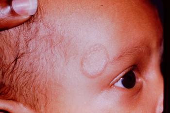
This lesion was treated in the primary care physician’s office. What treatment approach would you opt for?
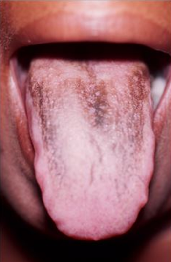
After a “swig” of Kaopectate, this patient noticed tongue discoloration. Is this coincidental, or is there a clinical connection?
From lacy and reticular and limited to the cheeks, this rash quickly covered the child’s body and transformed into a solid rash. Your dx?
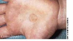
The location is unusual, but the appearance is characteristic. Your dx?
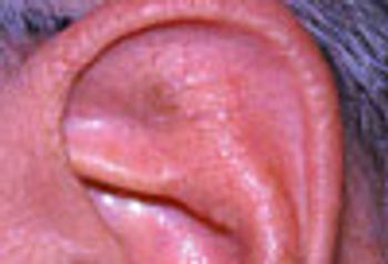
Skin lesions in the outer ear-the pinna, the concha, and the external auditory meatus-may be trivial or potentially malignant. Here we consider a flesh-colored swelling with a central white spicule; a tan papillary lesion; a fluctuant swelling; a red berry-like papule; an elevated pink lesion.
Benign skin lesions can arise in the outer ear as easily as in any other body part frequently exposed to the sun. Seborrheic keratosis may mimic malignant melanoma but is innocuous. Actinic keratosis is premalignant and should be excised, biopsied, and the site of excision monitored vigilantly.
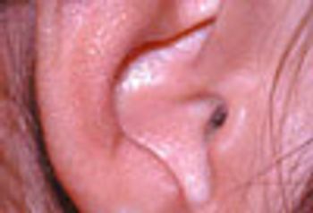
These innocuous lesions of the outer ear may arise spontaneously or after trauma or surgery. Both auricular seroma and pyogenic granuloma usually resolve satisfactorily after minor surgery, though they may recur.
Diagnostic challenge: Two case reports of easily treated and innocuous causes of lesions in the outer ear. Chondrodermatitis nodularis helicis is associated with long cellphone use. Verruca vulgaris is caused, like all other warts, by human papillomavirus.
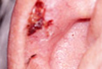
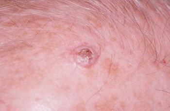
This rounded 0.5-cm ulcerated lesion with a slightly raised border between the helix and the antihelix had been present on an 85-year-old man’s ear for an unknown period.
An 87-year-old man experienced a scratchy throat and difficulty speaking, which cleared after taking over-the-counter throat lozenges.
Acanthosis nigricans is a nonspecific increase of the thickness of the prickle cell layer of the skin and most commonly seen in obese persons.
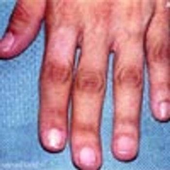
Shortly after awakening, a 17-year-old man experienced acute onset of severe pubic area pain.
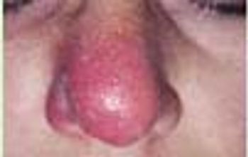
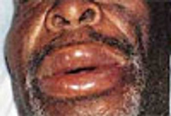
The nasal cellulitis that affects this 39-year-old woman began as right intranasal folliculitis.
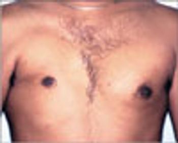
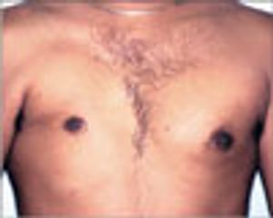
A 23-year-old man's right chest shows a common congenital muscle anomaly-partial absence of the pectoralis major muscle. The abnormality was noted during a routine preemployment physical examination. The clavicular origin seemed to be intact. There was no apparent decrease in shoulder internal rotation or adduction strength, and the patient had not noticed any shoulder weakness or limitation in motion.
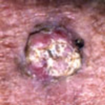
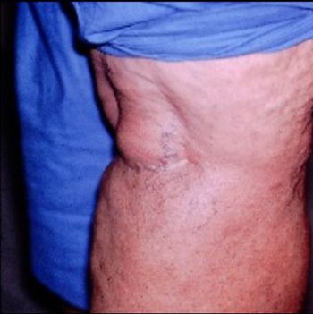
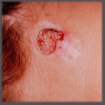
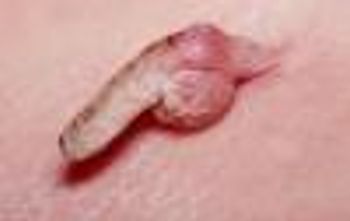
An 89-year-old man reported that this lesion began developing on his left forearm 11 days earlier. It is a keratoacanthoma, a rapidly growing but benign neoplasm that occurs predominantly on the extensor surfaces of the hands and forearms of white men over age 50.
Published: June 2nd 2008 | Updated:
Published: October 1st 2006 | Updated:
Published: September 14th 2005 | Updated:
Published: September 14th 2005 | Updated:

Published: September 14th 2005 | Updated:
Published: September 14th 2005 | Updated: