

News|Articles|November 17, 2005
Blue Rubber Bleb Nevus Syndrome
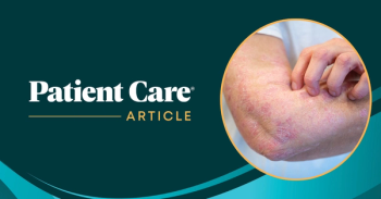
A 49-year-old man presented to the emergency department with hematemesis and 2 episodes of melena. Examination findings included resting tachycardia and melenic stool. Blood pressure was 95/50 mm Hg. Multiple raised, soft, bluish 0.3 to 1 cm lesions were noted on the trunk and extremities.
Advertisement
A 49-year-old man presented to the emergency department with hematemesis and 2 episodes of melena. Examination findings included resting tachycardia and melenic stool. Blood pressure was 95/50 mm Hg. Multiple raised, soft, bluish 0.3 to 1 cm lesions were noted on the trunk and extremities.
Fluids were given and the patient underwent an esophagogastroduodenoscopy (EGD). The EGD revealed multiple blood clots in the stomach and several reddish blue hemangiomas similar to those found on the patient's skin. Blood was actively oozing from one of these stomach lesions; endoscopic therapy with a thermal probe obliterated the bleeding site. The patient experienced no further bleeding. He was discharged from the hospital 2 days later.
Drs Klaus E. Mnkmller and Lucia C. Fry of the University of Alabama at Birmingham report that an association between cutaneous vascular nevi, intestinal lesions, and GI bleeding was first described in 1860; this constellation of findings was later named “blue rubber bleb nevus syndrome” to distinguish it from other cutaneous vascular lesions. A family history of this syndrome occurs infrequently; a few cases of autosomal dominant transmission have been reported.
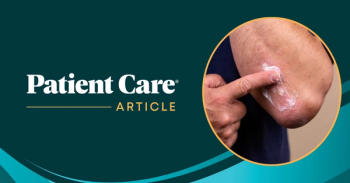
These distinctive lesions are blue, raised, wrinkled, and of variable size (from 0.1 to 5 cm). Characteristically, the contained blood can be emptied by direct pressure, leaving a wrinkled sac. Single or innumerable hemangiomas usually arise on the trunk, extremities, and face-but not on mucous membranes. Larger, disfiguring hemangiomas and darker, flat macules or papules also may develop.
The small bowel is the most commonly affected site; however, any part of the GI tract can be involved. Blebs in the colon most often are located distally. Occasionally, lesions have been found in the liver, lung, eye, CNS, and peritoneal cavity.
These lesions are best demonstrated by endoscopy; infrequently, they have been detected by barium or angiographic studies. Microscopically, they are cavernous hemangiomas that are composed of clusters of dilated capillary spaces.
Resection of the involved segment of bowel is indicated for patients with recurrent hemorrhage. Because these hemangiomas may involve the full thickness of the bowel wall, endoscopic laser coagulation can cause perforations and is therefore dangerous.
References:
FOR MORE INFORMATION:
- Gallo SH, McClave SA. Blue rubber bleb nevus syndrome: gastrointestinal involvement and its endoscopic presentation. Gastrointest Endosc. 1992;38:72-76.
- Yamada T, Alpers DH, eds. Textbook of Gastroenterology. 3rd ed. Philadelphia: Lippincott Williams & Wilkins; 1999.
Newsletter
Enhance your clinical practice with the Patient Care newsletter, offering the latest evidence-based guidelines, diagnostic insights, and treatment strategies for primary care physicians.
Advertisement
Related Content
Advertisement


Latest CME


Advertisement
Advertisement
Trending on Patient Care Online
1
Long-Term Data Support Sustained Bimekizumab Response in Hidradenitis Suppurativa
2
First Oral Film Treatment for Erectile Dysfunction in Men Gains FDA Approval
3
Weekly Dose Podcast: New Obesity Data, Insulin Guidance, and Mental Health Screening
4
Topline Phase 2 Data Show Roflumilast Cream Improves Atopic Dermatitis in Infants as Young as 3 Months
5