

|Articles|May 1, 2008
Anal Pain: Office Diagnosis and Treatment
Author(s)Michael E. Friscia, MD, Robert D. Fry, MD
Patients almost always believe that their anorectal problems are caused by hemorrhoids, regardless of the nature of their symptoms. They are often dismayed when we insist that they must come to the office for an examination before we can prescribe any treatment.
Advertisement
FIND OUT MORE
Patients almost always believe that their anorectal problems are caused by hemorrhoids, regardless of the nature of their symptoms. They frequently phone and ask us to prescribe a medication for their "painful hemorrhoids," and they are often dismayed when we insist that they must come to the office for an examination before we can prescribe any treatment.
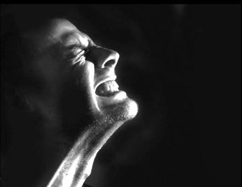
While it is certainly understandable that our patients should make such an assumption, it is all the more important for us, as health care providers, to remember that anal pain frequently has a nonhemorrhoidal cause. In fact, 95% of patients who complain of anal pain have one of the following 3 conditions: anal fissure, thrombosed hemorrhoid, or anorectal abscess. In this article, we discuss the diagnosis and treatment of these conditions, as well as other, less common causes of anal pain.
FISSURE
An anal fissure is simply a tear in the anoderm. In men and most women, the tear, or ulcer, occurs very close to the posterior midline of the anal canal. The linear injury is typically caused initially by the passage of a hard stool, resulting in pain with defecation that can be accompanied by bleeding. The pain causes sphincter spasm and anal hypertonia, and thus begins a vicious circle of pain, spasm, and constipation. This syndrome is associated with relative ischemia of the posterior midline of the anal canal, an area with a relative paucity of blood vessels.1
Often by the time patients arrange an appointment, what started as a tear has become an ischemic ulceration. At this point, the adjacent perianal skin may be edematous, forming a "sentinel pile"-the hallmark of a chronic fissure that will likely need surgical treatment. This swollen epithelial tag may resemble an external hemorrhoid. The anal papilla at the cephalad edge of the anal fissure may also become enlarged and hypertrophied as a result of the chronic inflammation. Thus, the classic triad of a chronic anal fissure is the fissure itself, accompanied above by a hypertrophied anal papilla and below by a sentinel skin tag.
Diagnosis. You can easily diagnose an anal fissure by carefully spreading the buttocks apart and observing the linear ulceration in the anal canal. At this point, further examination of the anorectum with either a finger or instrument is unnecessary and can be quite painful for your patient. Up to 10% of fissures occur in the anterior midline (particularly in women). Fissures that are not close to the midline suggest other processes that could result in anal or perianal ulceration, such as Crohn disease, syphilis, herpes, and carcinoma.
Treatment. Your patient may well express some anxiety about the pain and bleeding associated with an acute anal fissure. For this reason, reassurance is beneficial-emphasize that most fissures heal spontaneously with nonoperative treatment. Standard treatment includes stool softeners, such as methylcellulose or dietary fiber. Warm sitz baths seem to provide comfort and may relax the anal spasm and provide gentle anal cleansing following bowel movements.
Topical therapy. Topical corticosteroids and suppositories are ineffective and should be avoided; topical anesthetics are popular, but we do not routinely recommend their use to our patients. Because ischemia has been implicated in the propagation of a chronic fissure, several topical smooth muscle relaxants have been shown to be effective, probably by both lowering resting anal pressure and promoting vasodilatation. We find that topical diltiazem (2% gel) may relieve the pain and spasm and promotes healing. Topical nitroglycerin (0.2% cream applied twice daily) has been used with success; however, we have found that recurrence is common and patients often complain of headaches that accompany its use.
When surgery is needed. If the fissure does not heal or if the pain does not diminish substantially over the next 2 to 3 weeks, a more aggressive approach is justified. In addition, the presence of a sentinel pile and a hypertrophied anal papilla implies that the fissure is a chronic problem that is unlikely to respond to nonsurgical treatments (
The internal anal sphincter resides immediately beneath the anoderm and thus actually forms the base of a deep fissure. The internal sphincter becomes relatively rigid in a patient with a chronic anal fissure, a condition that seems to be associated with fibrosis of a portion of the sphincter. This rigidity of a portion of the internal sphincter can be appreciated by digital examination of the anal canal, but the pain associated with the fissure usually mandates that a local anesthetic be administered before such an examination. Relief of this rigidity is provided by internal anal sphincterotomy, a procedure that is nearly always curative.
Preferred technique. Most surgeons agree that the operation of choice for a fissure that will not heal is a lateral subcutaneous internal sphincterotomy, which can easily be performed in the outpatient setting using local anesthesia. Here's how we do it:
With the patient in the prone jackknife position, we infiltrate the perianal tissues on one side of the anus with 15 mL of 0.25% bupivacaine with epinephrine. We always include the area adjacent to the fissure in the anesthetic field so that the anus can be dilated comfortably. Next, the proctoscope is inserted. Careful proctoscopy is performed to ensure that the patient has no other pathological anorectal condition.
We then insert a Hill-Ferguson anoscope into the anal canal, which stretches the anal sphincters. With the sphincters stretched by the instrument, the intersphincteric groove is easily palpable, as is the abnormally fibrotic portion of the internal sphincter that causes the fissure to persist. With the left index finger in the anus, a No. 11 blade scalpel is inserted into the intersphincteric groove, with the blade parallel to the internal sphincter (Figure 2 [
Postoperative care. Our patients have surprisingly little discomfort after this procedure. We prescribe acetaminophen and ask them to continue sitz baths for a few days. We re-examine the patient in 2 weeks to verify healing. The published success rate for this procedure is greater than 90%, and complications such as incontinence are quite rare.2THROMBOSED HEMORRHOIDDiagnosis. The second most common cause of anal pain is a thrombosed external hemorrhoid. The patient reports pain of sudden onset that is associated with an anal mass. The diagnosis is immediately apparent when you see a swollen, dark-colored mass located at the anal verge. This is a clot in the external hemorrhoidal vein. The key here is to understand the time course of this process: the pain is most intense during the first 24 to 48 hours after the clot forms, and then it resolves rapidly, even with no treatment.
Treatment. Because surgical treatment of a thrombosed hemorrhoid is usually painful, we try to determine whether the procedure will cause more pain than conservative management. If the patient's pain is beginning to diminish, we usually recommend medical therapy with topical corticosteroids, warm soaks, and stool softeners.
Only if the patient has severe pain do we recommend excision. Although we prefer to perform the procedure in the operating room, it can be accomplished in the office with the aid of a local anesthetic (0.25% bupivacaine). The bupivicaine is infiltrated under and around the hemorrhoid, and the entire hemorrhoid, consisting of skin and thrombosed vein, is excised in an elliptical fashion. Electrocautery can be used for hemostasis. It is acceptable to leave the wound open and allow healing to occur by secondary intention, but we often approximate the skin edges with a running 3-0 absorbable suture. The entire hemorrhoid must be completely excised, because simply evacuating the thrombus often results in reaccumulation of the thrombus.
Patients usually have significant discomfort after the excision of a thrombosed hemorrhoid, so we recommend a leave from work for several days. The postoperative care is similar to that described for sphincterotomy.
ANORECTAL ABSCESS
The third common cause of anal pain is an anorectal abscess. This condition is the most subtle, and yet the most dangerous, of the three. Even if a fissure or thrombosed hemorrhoid remains untreated, it will likely resolve; it almost never becomes worse. In contrast, an untreated abscess can extend to adjacent areas and, in some instances, can be life-threatening, particularly in immunocompromised patients.
Origins and types of abscesses. Most anorectal abscesses originate in the anal glands at the base of the anal crypts. These glands are located between the internal and external anal sphincters. Thus, the origins of anorectal abscesses are cryptoglandular, and since the process arises in the space between the internal and external sphincters, most abscesses originate as intersphincteric abscesses. The intersphincteric plane is a conceptual space rather than a real one, and abscesses arising in the location must find egress as they expand (
Diagnosis. When a patient complains of anal pain, and the examination does not reveal a fissure or a thrombosed hemorrhoid, the cause is most likely an anorectal abscess. The perianal area should be carefully inspected and palpated. A perianal abscess is usually obvious: a tender, indurated lump located at or just distal to the anal verge. Only advanced abscesses are fluctuant, and pain may precede the development of induration. If pressure over the skin surrounding the anus or the buttocks does not elicit pain or reveal induration, a gentle digital anal examination is required. Because virtually all anorectal abscesses are cryptoglandular in origin, palpation of the anal canal should reveal tenderness and (usually) induration caused by the infection harbored in the anal crypt.
In rare circumstances, the infectious process will extend upward toward the pelvis. This can be detected by tenderness and induration in the rectal wall overlying the "high" abscess.
If the suspected abscess cannot be confirmed by physical examination, including a careful digital rectal examination, a CT scan may be required. However, we obtain CT scans for this condition only rarely. The diagnosis can be accurately made by physical examination in more than 95% of patients with anorectal abscesses.2Treatment. The only appropriate treatment of an anorectal abscess is incision and drainage. Cryptoglandular abscesses do not resolve with antibiotic treatment. In general, reserve antibiotics for immunocompromised patients and for those who have significant cellulitis associated with the abscess.
Although it is possible to accomplish incision and drainage in the office, the procedure is uncomfortable for the patient and adequate exposure is difficult to achieve in this setting. In addition, if the crypt containing the gland from which the infection arose ("the offending crypt") can be detected, the crypt and gland can be eradicated at the same time that the abscess is drained; this significantly reduces the likelihood of fistula formation. For these reasons, we treat anorectal abscesses in the controlled environment of the operating room.
Some surgeons use a local anesthetic, but we prefer either a general or spinal anesthetic. Injecting the tender, inflamed tissue associated with the abscess is extremely uncomfortable for the patient. In addition, it is very difficult to achieve sufficient relaxation with a local anesthetic to permit an adequate examination. In fact, infiltrating the anal tissues with a local anesthetic causes edema that makes it more difficult to find the source of the abscess.
After adequate anesthesia has been achieved, the patient is placed in the prone flexed position and careful examination with an anoscope is performed. Pressure placed over the abscess may produce pus from the crypt containing the infected gland. A crypt hook is inserted in the abscess, and an incision with electrocautery over the crypt hook results in division of skin and a portion of internal sphincter and allows unroofing of the abscess. The opening over the abscess is further enlarged by excising a 1-cm disk of skin to allow adequate drainage of the pus. We do not pack the cavity with gauze, nor do we use drains. If the crypt of origin is detected and the abscess drained as described (this is actually a fistulotomy), the risk of recurrent infection is less than 5%.3
If the offending crypt cannot be detected, the abscess is drained by making a cruciate incision over the abscess as close to the anal verge as possible. The edges of the incision are then excised to create an opening of about 1 cm in diameter. This procedure effects a cure in about 50% of patients.3
In half of the patients treated only by incision and drainage, a chronic fistula forms. The internal opening to the fistula is the crypt containing the chronically infected anal gland, and the external opening is the site where the incision was made to drain the abscess. For this reason, we place the incision as close to the anal verge as possible so that a fistula that may later form will be as short as possible.
OTHER CAUSES
While fissures, thrombosed hemorrhoids, and anorectal abscesses account for the vast majority of cases of anal pain, keep in mind that other, less common causes may be responsible, particularly in immunocompromised patients.
Herpes. Anorectal herpes simplex lesions can be quite painful. The diagnosis can be difficult initially because severe pain may precede the appearance of the typical clustered vesicles surrounded by a red areola. The lesions can occur on the rectal mucosa, anoderm, and perianal skin. The diagnosis is confirmed by viral culture of fluid from a vesicle. Remind your patient that these lesions are highly infectious. Oral acyclovir or valacyclovir shortens the duration of symptoms and reduces the frequency of episodes.4 Recurrence is, of course, the rule.
Syphilis. Multiple fissures or soft, symmetrical lateral fissures point to a diagnosis of syphilis. These lesions, which can be quite painful, often resemble poured wax after it has cooled. The characteristic appearance coupled with a positive rapid plasma reagin test are sufficient to make the diagnosis. If the diagnosis is still in question, obtain serum from the base of the lesion and order a fluorescent treponemal antibody-absorption test. The mainstay of treatment for syphilis continues to be long-acting penicillin preparations.
Infectious proctitis. An intense proctitis may result from infection with Chlamydia trachomatis or Neisseria gonorrhoeae, which can lead to anorectal pain and tenesmus. Send swab samples for gonococcal culture; negative results suggest chla- mydial infection. Rising titers of antibody to C trachomatis may help pin down the diagnosis. Doxycycline can be used to treat chlamydial infections; however, gonococcal infections require consultation with local health officials because of widespread antibiotic resistance.
CLINICAL HIGHLIGHTS
• In men and most women, anal fissures occur very close to the posterior midline of the anal canal. Fissures that are not close to the midline suggest other processes that could result in anal or perianal ulceration, such as Crohn disease, syphilis, herpes, and carcinoma.
• Most fissures heal with conservative treatment, including stool softeners, warm sitz baths, and topical smooth muscle relaxants. If the fissure does not heal or if the pain does not diminish substantially over 2 to 3 weeks, a more aggressive approach is justified; internal anal sphincterotomy is nearly always curative.
• The pain of a thrombosed hemorrhoid is usually most severe during the first 24 to 48 hours after the clot forms, and then it resolves rapidly. If the patient continues to have severe pain-despite conservative treatment with topical corticosteroids, warm soaks, and stool softeners-excision is recommended.
• A perianal abscess is usually obvious: a tender, indurated lump located at or just distal to the anal verge. Only advanced abscesses are fluctuant, and pain may precede the development of induration.
• The only appropriate treatment of an anorectal abscess is incision and drainage. Reserve antibiotics for immunocompromised patients and for those who have significant cellulitis associated with the abscess.
References:
REFERENCES:
1. Schouten WR, Briel JW, Auwerda JJ. Relationship between anal pressure and anodermal blood flow. The vascular pathogenesis of anal fissures. Dis Colon Rectum. 1994;37:664-669.
2. Metcalf A. Anorectal disorders. Five common causes of pain, itching, and bleeding. Postgrad Med. 1995;98:81-84, 87-89, 92-94.
3. Bastawrous AL, Cintron J. Anorectal abscess and fistula. In: Cameron JL, ed. Current Surgical Therapy. 8th ed. Philadelphia: Mosby; 2004:256-261.
4. Leone PA, Trottier S, Miller JM. Valacyclovir for episodic treatment of genital herpes: a shorter 3-day treatment course compared with 5-day treatment. Clin Infect Dis. 2002;34:958-962.
Advertisement
Related Content
Advertisement


Advertisement
Advertisement
Trending on Patient Care Online
1
Nearly 18 Million Middle-Aged US Women Overdue for Cancer Screening, Study Finds
2
Tirzepatide and Emerging OSA Pharmacotherapy Expand the Treatment Toolkit, With Ashesha Mechineni, MD
3
HNS Tied to Reductions in 8 of 9 MACE Outcomes in Late-Breaking OSA Analysis: SLEEP 2026
4
When CPAP Fails: Expert Discussion on How to Counsel Patients With OSA
5