

|Articles|April 26, 2013
Pain Problems-A Photo Essay
Author(s)Leo Robert
Lofgren syndrome, lumbar disk herniation, suppurative appendicitis with rupture, rotator cuff impingement syndrome, aortic dissection.
Advertisement
A 33-year-old woman presented with frontotemporal headaches and neck pain. Later, she presented to the ED with a worsening headache now accompanied by nausea, vomiting, ankle pain with swelling, and fever. A new rash on the anterior aspect of her lower extremities (below) appeared as erythematous, tender patches, 3 to 4 cm in size. Biopsy of lung tissue was significant for granulomatous disease, and a diagnosis of
Image courtesy of Mohenish Singh, DO, Brandon Hill, MD, and James McDonald, MD.
Click here for the next image
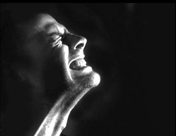
This patient has
Image courtesy of David Della-Giustina, MD and Bradford A. Kilcline, MD.
Click here for the next image
Abdominal pain in older patients is a medical Pandora’s box-some die, many need surgery, and the cause often remains unknown. An 84-year-old woman presented with nausea, vomiting, and constant lower abdominal pain. Abnormal urinalysis results and a normal WBC count were consistent with a urinary tract infection. This helical CT scan suggested perforating appendicitis. Surgical exploration revealed
Image courtesy of Carolyn J. Sachs, MD, MPH.
Click here for the next image
A 55-year-old right-handed man had a constant dull ache in his right shoulder that worsened when he steered his car or elevated his arm. The pain radiated to his neck and upper right arm. In the “empty can” test (below), a patient abducts both arms 90° laterally, then adducts them 20° to the frontal plane, with thumbs turned down; the examiner resists further abduction to test strength and watches for signs of pain. The man most likely had
Image courtesy of Edward J. Shahady, MD, Willis Paull, PhD, and Seth Smith, PharmD.
Click here for the next image
A 57-year-old woman presented with severe chest pain and general malaise. She had a history of hypertension. A frontal upright radiograph of the chest showed a prominent mediastinum at the upper limits of normal. The aorta was ectatic. A CT scan without contrast through the ascending aorta revealed a linear area of fluid within the ascending aorta (B, arrow). The fluid represents blood in the displaced intimal wall of the ascending aorta, which is diagnostic of
Image courtesy of William Yaakob, MD and Stephen Schabel, MD.
Advertisement
Related Content
Advertisement


Advertisement
Advertisement
Trending on Patient Care Online
1
Semaglutide Boosts Quality of Life in T2D and CKD, FLOW Trial Analysis Finds
2
5 FDA Decisions for Primary Care to Know from May 2026
3
Shared Decision-Making and MHT Safety in Women with VMS
4
FDA Issues Complete Response Letter for Cingulate ADHD Drug CTx-1301
5