
Opinion|Videos|July 7, 2025
Turning Images Into Action: Identifying Suspicious Lesions With Confidence
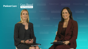
Panelists discuss that decisions to forgo biopsy often weigh patient risk and lesion characteristics with noninvasive tools offering valuable reassurance; while familiarity with artificial intelligence (AI) aids varies, these technologies—used judiciously and alongside clinical expertise—can boost diagnostic confidence, though limitations and integration challenges remain.
Advertisement
Episodes in this series
Sometimes, the decision not to biopsy a lesion comes down to weighing the risks and benefits, especially with older or medically fragile patients. If a lesion appears low-risk and the patient might not tolerate a biopsy well, clinicians may choose to monitor rather than intervene immediately. Biopsies remain the gold standard for diagnosis, but they can sometimes complicate care or create unnecessary patient discomfort. In these cases, tools that help rule out malignancy or provide additional reassurance without invasive procedures are especially valuable. This approach can reduce patient anxiety and avoid unnecessary medical risks when clinical judgment supports it.
Familiarity with skin cancer detection tools varies widely among practitioners. While some have integrated such technologies into their practice, many remain curious but uncertain about their practical utility. Educational resources from dermatology organizations provide excellent training materials to help bridge this gap. Confidence in diagnosing skin cancer tends to grow with experience and validation, and AI tools can enhance this by providing immediate feedback and support. This allows providers to detect cancers earlier and expand their diagnostic capabilities. Importantly, these tools are meant to supplement—not replace—clinical expertise, fostering better outcomes by increasing diagnostic confidence and reducing unnecessary referrals.
Certain lesions are less suitable for AI-based diagnostic tools, particularly those located on sensitive areas such as eyes or lips or lesions with heavy crusting. These devices are designed primarily to assess suspicious lesions with atypical cells, especially common skin cancers such as basal cell carcinoma, squamous cell carcinoma, and melanoma. The proper use and understanding of a device’s limitations are essential to avoid misapplication. Additionally, barriers such as cost and workflow integration continue to challenge adoption in clinical settings. Still, with appropriate training and system integration, these technologies can enhance primary care skin cancer detection and reduce referrals, benefiting both patients and providers.
Advertisement
Related Content
Advertisement


Advertisement
Advertisement
Trending on Patient Care Online
1
Bepirovirsen and the Push to Close Hepatitis B Care Gaps
2
FDA Grants Fast Track Designation to HTX-001 for Symptomatic Nonobstructive Hypertrophic Cardiomyopathy
3
Noninvasive Testing and the Colorectal Cancer Screening Gap, With Mark Fendrick, MD
4
CorVista System Receives FDA Clearance for Noninvasive PCWP Test
5