Articles by Samer Alkhuja, MD
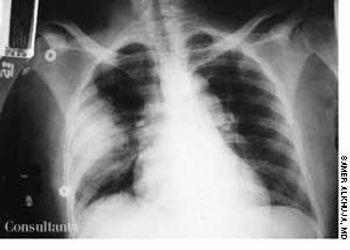
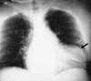
A febrile 65-year-old woman who had suffered a new-onset seizure was brought to the emergency department. The patient-a cigarette smoker-was not coughing and had neither chest pain nor a significant medical history. Her temperature was 39.4°C (103°F). She had nuchal rigidity and Kernig's and Brudzinski's signs of meningeal irritation. Lung auscultation revealed signs of right middle lung consolidation. Her white blood cell count was 1,200/µL. A chest film, seen here, showed a masslike density in the right midlung.
A 35-year-old man, a smoker, had right pleuritic pain, productive cough, and fever for 3 days. His pulse rate was 107 beats per minute; respiratory rate, 14 breaths per minute; blood pressure, 136/80 mm Hg; and temperature, 37.7°C (99.9°F). There were signs of right upper lobe consolidation. Laboratory studies showed hyponatremia. Chest films showed a homogeneous density in the right upper lobe.
Except for a fever of 1 month's duration, a 28-year man had no other complaints. He said he used to smoke marijuana but never used intravenous drugs. His temperature was 39.4°C (103°F), but no other abnormalities were noted on physical examination.
A 49-year-old woman, severely obese but otherwise healthy, appeared for a preemployment medical examination. She neither smoked cigarettes nor drank alcohol. She had no respiratory problems and recalled no family history of such. A baseline mammogram taken 4 years earlier showed no abnormalities, and the patient was not under care for any medical condition. Results of physical examination were normal, except for the obesity-which made it difficult to determine breast masses with confidence.
A 52-year-old man from Bangladesh had suffered from pleuritic pain for 1 week. He had never had tuberculosis and-except for being a cigarette smoker-had no notable medical history. The only remarkable findings were a temperature of 37.5°C (99.5°F) and anterior tenderness over the right lower rib cage. Laboratory test results were normal. A tuberculin test with 5 TU of purified protein derivative produced positive results, with a 15 × 17-mm induration.
Presenting symptoms of this 42-year-old man were left pleuritic pain and severe dyspnea while climbing stairs. He had a 2-year history of exertional dyspnea but had not sought medical advice. The patient's pulse was 123 beats per minute; respiratory rate, 45 breaths per minute; blood pressure, 80/45 mm Hg; and temperature, 37.3°C (99.1°F). Chest examination revealed hyperresonance with absence of breath sounds over the left hemithorax and wheezing over the right lung.
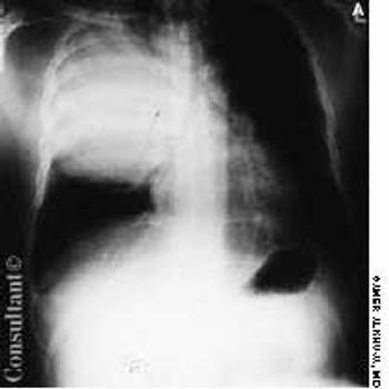
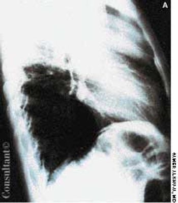
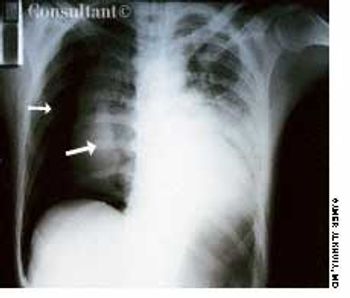
During the assessment of an 83-year-old man who had an infected arteriovenous graft, chest films showed a masslike density in the lower right hemithorax.
A 40-year-old woman with AIDS had been feverish for the past 24 hours and had a nonproductive cough. She had smoked one pack of cigarettes daily for 20 years.
Fever of 1 month's duration was this 28-year-old man's only complaint. He had smoked marijuana in the past but denied intravenous drug use. His temperature was 39.4°C (103°F), but no other abnormalities were noted on physical examination.
While watching TV, a 32-year-old man experienced acute right-sided pleuritic pain and was taken to the emergency department. He was seropositive for HIV but had never had Pneumocystis carinii pneumonia (PCP) and was not taking aerosolized pentamidine. Physical examination revealed hyperresonance with significantly decreased breath sounds over the right hemithorax.
For the past 2 days, a 30-year-old man had experienced scant hemoptysis. He had also lost a significant amount of weight-5 kg (11 lb)-over the last 2 months. The patient, a recent emigrant from Ecuador, had no history of tuberculosis (TB) or of exposure to this disease, and he had not been skin tested with purified protein derivative (PPD). He denied exposure to risk factors for HIV infection.
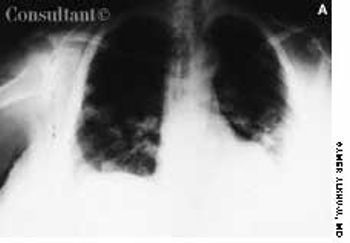
A previously healthy 51-year-old man presented with weight loss and poor appetite of 2 months' duration. He was heterosexual and had many sexual partners. Except for a temperature of 38.3°C (100.9°F) and left basal rhonchi, results of physical examination were normal. A chest radiograph and CT scan, as seen here, showed large cavitary lesions in the lower left lobe.
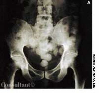
A 2-year-old boy was hospitalized because of acute abdominal pain. He had no other symptoms. The child's temperature was 37.3°C (99.1°F). He was irritable and had generalized abdominal tenderness. A stool test was positive for blood. Results of laboratory investigations were normal.
For the past 3 months, a 66-year-old man has suffered fatigue and loss of appetite and weight. He was not coughing, nor had he experienced fever, chest pain, or hemoptysis. He had no history of notable respiratory disease, and he was not aware of having had tuberculosis (TB).
A 54-year-old woman with a history of hypertension presented with a worsening headache and a left hemisensory defect. A CT scan of her head without contrast showed a right parietal hemorrhage with spreading edema; the masslike effect caused shifting of the midline to the contralateral side. The patient gradually became comatose and required intubation for airway protection. Intravenous corticosteroids were administered to decrease the effect of the lobar hemorrhage. Fever developed 3 days after admission.

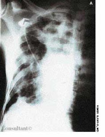
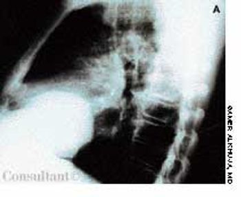
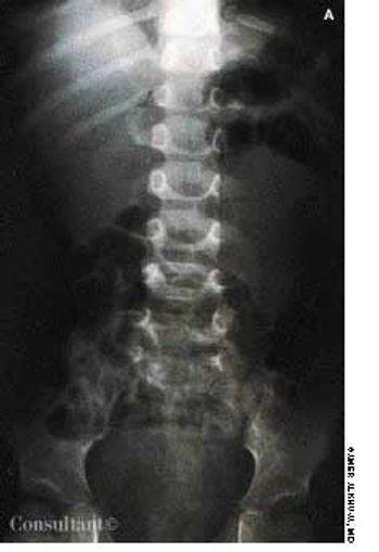
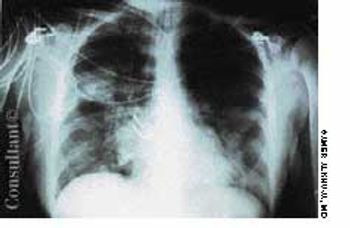
A 70-year-old man with a history of peptic ulcer disease presented with a 1-dayhistory of epigastric pain. Abdominal examination revealed mild epigastrictenderness. A pneumoperitoneum was discovered on a chest film (A), and aleft decubitus chest film (B) confirmed this diagnosis.


