Articles by Matthew C.k. Choi, MD
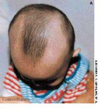
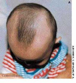
Concerned about their son's progressive hair loss during the last 6 months, his parents brought the 2-year-old into the office. The clinical appearance of hair loss extending in a band configuration around the temporal-occipital scalp margin confirmed the diagnosis of ophiasis.
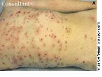
A pruritic, erythematous rash developed in a 6-year-old boy over the previous 5 days. The rash erupted in crops; the lesions appeared initially as rose-colored macules, progressed rapidly to papules and vesicles, and finally crusted. The distribution of the lesions-with the greatest concentration on the trunk-is typical of chickenpox.
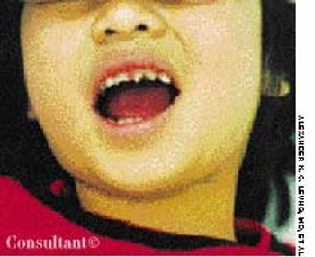
These notched upper central incisors were noted in a 3-year-old girl. There was no history of traumatic injury to her mouth. The youngster was able to sweat normally, and no other evidence of ectodermal dysplasia was present.