
Charles E. Crutchfield III, MD
Advertisement
Articles by Charles E. Crutchfield III, MD
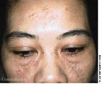
Loss of pigmentation was noted around the left eyeof a 49-year-old man-the same eye in which hehad a detached retina. In addition, the patient'shearing was impaired on that side. These findingswere consistent with the diagnosis of Alezzandrinisyndrome, the major manifestations of which includeunilateral degenerative retinitis, ipsilateral facial vitiligo,poliosis of the eyebrows and eyelashes, and ipsilateralhearing deficits.
A 51-year-old man presented with red, mildly pruritic papulonodules that had erupted on his face approximately 5 weeks earlier. The clinical appearance suggested cutaneous lymphoid hyperplasia.
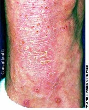
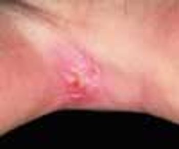
Extremely itchy, crusting nodules appeared on the arms and legs of a 42-year-old woman who was undergoing renal dialysis.
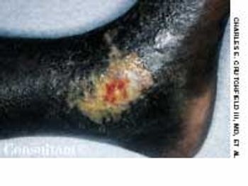
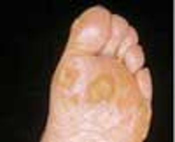
Cutaneous manifestations develop in approximately 30% of persons with diabetes. Premature atherosclerosis is a common complication of the disease that can cause peripheral infarction, ulceration, and necrosis.

This rare condition affects both men and women. The average age at onset is 53 years. The lesions are deep brownish red to purple papules, nodules, and plaques. Blisters and ulcers also can occur.
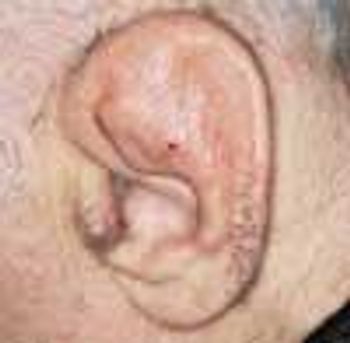
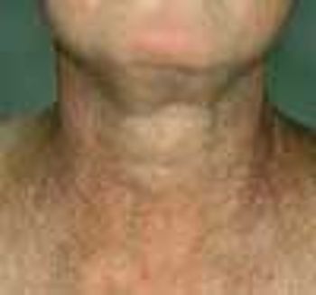
Relapsing polychondritis, as manifested in one of this 55-year-old man's deformed ears, is a rare, chronic, autoimmune inflammatory disease of cartilaginous structures. The disease also affected the cartilage in this patient's nose, which is the second most common site of involvement. Tissues of the joints, eyes, and blood vessels as well as the trachea and the bronchial tree may also be affected and destroyed.
A 39-year-old man who has sickle cell disease suffers with chronic ankle ulcers typical of the disorder. Ulcerations occur in approximately 50% of persons who are homozygous for sickle cell disease.
A 67-year-old woman sought medical treatment for the persistent, large “blackheads” on her face. Examination revealed diffusely thickened and yellowed skin with deep furrows. The periorbital and malar areas were studded with large open comedones.
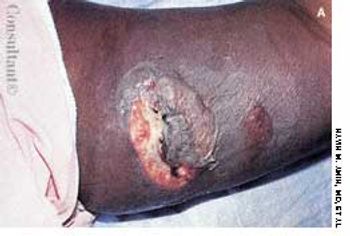
A 61-year-old woman who was receiving dialysis for diabetes-associated end-stage renal disease was hospitalized for care of an abdominal wound that had been debrided and closed. At this time, the patient had several large, indurated, red plaques with central, stellate, black eschars on her abdomen, left buttock, and legs. An early focus of ulceration was noted superior to the stapled incision.
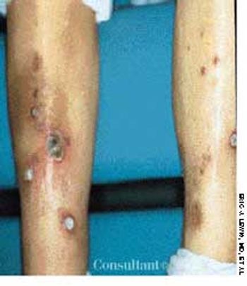
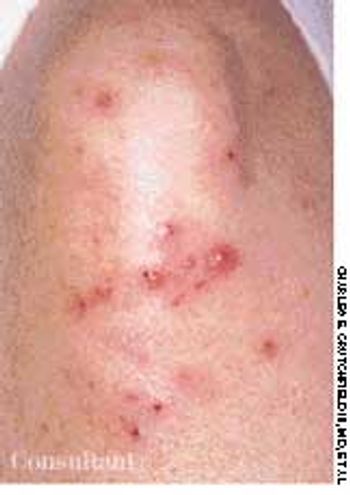
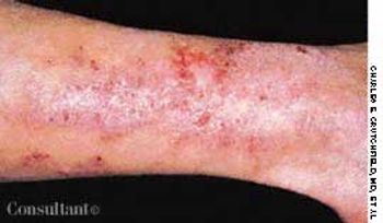
A 63-year-old woman who was on long-term hemodialysis because of diabetic end-stage renal disease had a 7-month history of waxing and waning papules and plaques on the front of both legs. The asymptomatic multiple, discrete, slightly erythematous, round to oval lesions ranged from 5 mm to 3 × 4 cm. Several had heaped-up borders and contained central crust and keratotic debris; others were superficial ulcers with central eschars. The lesions improved only slightly following twice-daily application of a superpotent topical corticosteroid preparation.
A 55-year-old-man complained of joint stiffness and red, mildly tender plaques on his fingers. He had recently sustained a trauma to the hand while at his job as a fish handler. The condition was diagnosed as erysipeloid-a skin infection caused by the gram-positive bacillus Erysipelothrix rhusiopathiae.
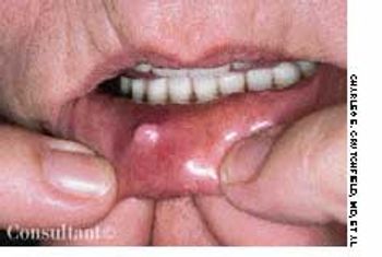
The painless lump on this 30-year-old woman's lip is a mucous cyst, or mucocele-the result of mucus retention within a salivary gland due to trauma or obstruction of a duct. This asymptomatic, dome-shaped, translucent, blue-white cyst is usually located on the inner surface of the lower lip or on the floor of the mouth.
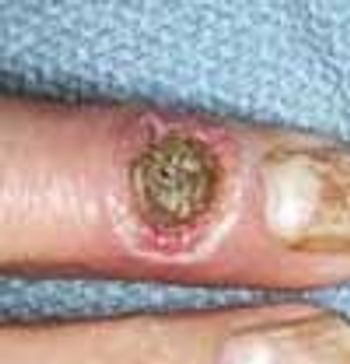
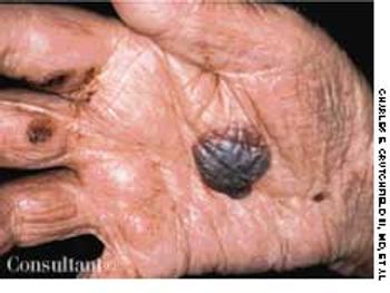
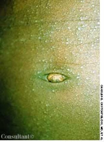
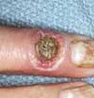
A 16-year-old boy removed a small sliver of wood from the palm of his hand with a pocket knife. However, the fledgling “surgeon” created a small puncture wound during this operation. Over the next month, a small, moist, friable papule grew at the site.
A 47-year-old woman from Southeast Asia presented with erythematous, asymmetric, anesthetic, sharply marginated plaques on her lower arms and hands. Because this patient had emigrated from a tropical climate, leprosy was suspected.
This lesion erupted on a 58-year-old man's right palm, and several tense bullae also were visible on the trunk. The patient complained of mild pruritus. He had no history of similar lesions. A routine skin biopsy was performed, and the diagnosis of hemorrhagic bullous pemphigoid was made.
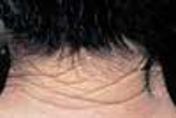
During a routine skin cancer screening, yellowed, thickened, leathery skin was noted on the posterior neck of a 73-year-old retired construction worker. Colloquially, this condition is called “sailor's skin” or “farmer's skin” and is seen in persons who have had long-term exposure to the sun. It is known clinically as cutis rhomboidalis nuchae, because the well-defined furrows in the skin resemble an irregular rhomboidal pattern.
An otherwise healthy 4-year-old boy was brought for evaluation of a mildly pruritic rash, which had been present for approximately 8 months. The developmental history of the eruption was equivocal, and the child's mother reported no aggravating or ameliorating factors.
A 41-year-old man sought treatment of the rough, intensely itchy patch on the dorsum of his left foot and lateral ankle. There were no other similar plaques on his body.
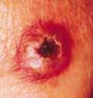
A 17-year-old high school athlete was anxious about this new “mole” that appeared on the heel of his right foot. His concern was prompted by the recent diagnosis of melanoma in his aunt.

An otherwise healthy 34-year-old woman was concerned because of the abrupt onset of rapid hair loss, accompanied by scaling of the underlying skin. The disorder had begun 3 months earlier, and the right parietal and temporal areas were now red and swollen and had adherent scale. An antinuclear antigen titer was negative. Biopsy revealed changes consistent with lichen planus of the scalp, also known as lichen planopilaris and lichen planus follicularis.
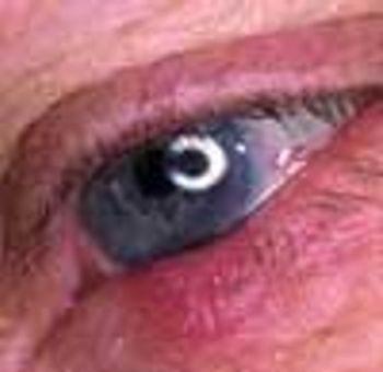
A mildly itchy, tender “red bump” on his eyelid concerned a 68-year-old man. Two years earlier, a basal cell carcinoma, which started as a “red bump,” had been removed from his cheek. This lesion had been present for 2 weeks; the patient noted a small amount of discharge at the site in the mornings.
A 44-year-old woman was being seen regularly for skin manifestations of systemic lupus erythematosus (SLE). During a routine visit, blotchy erythema and hyperpigmentation were noted on the normally exposed areas of her neck and upper chest; the submental area was spared. Close examination revealed fine telangiectases and poorly marginated hyperpigmented and hypopigmented macules.
A 63-year-old farmer first noticed multiple rough bumps on his hands and feet at least 20 years before pointing them out to his physician. A diagnosis of arsenical keratoses was made after the patient reported that as a child he had worked on his family's potato farm, where a commonly used pesticide, “Paris Green,” was applied to the plants. The active ingredient in this pesticide was inorganic arsenic.
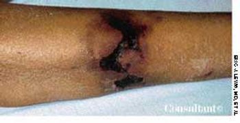
A 42-year-old woman was hospitalized with an increasingly painful, ulcerated swelling of the right lateral thigh. The patient reported that while cleaning out her attic, she suddenly experienced an excruciating burning sensation in her thigh. Immediately, she rubbed the area with ice, which provided partial relief. An ulcer developed 24 hours later and began to enlarge.
A 40-year-old farmer had been complaining for 3 weeks of a tender, red, itchy, scaling plaque with papulopustules on one knee. A potassium hydroxide examination of the scale revealed fungal hyphae.
A 40-year-old man presented with tiny, intensely pruritic vesicles on the knees, legs, buttocks, elbows, and scalp.
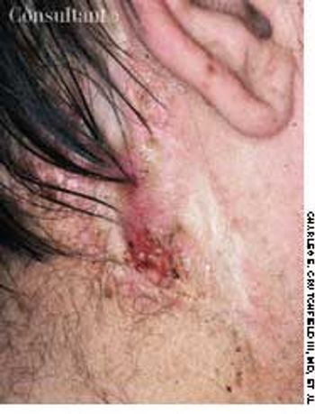
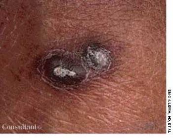
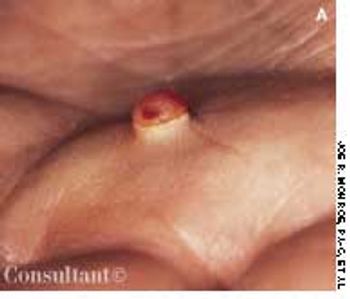
An ulcerating nodule on his left pre-auricular region worried a 64-year-old man.
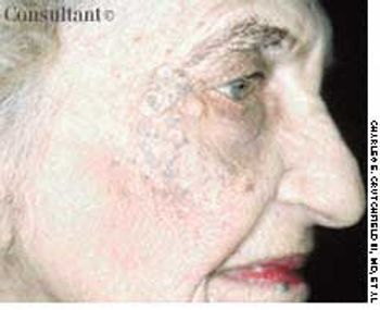
A 32-year-old woman became concerned about the numerous bumps on her upper face, which had been increasing in number for many years. Examination revealed clusters of 2- to 3-mm lesions-mostly monomorphic, flesh-colored papules lateral to the eyebrows and on the central forehead, inferior-medial eyelids, and upper cheeks. Some of the papules had a slight pink or yellow-brown appearance, and poorly demarcated brown patches (melasma) were noted on the cheeks.
A scaling, red, fissured area between digits 4 and 5 on her right hand sent a 33-year-old woman to her physician. Diagnosis of interdigital Candida was confirmed by a potassium hydroxide evaluation of material from the site.
Advertisement
Latest Updated Articles
 A Case of Giant Colonic Diverticulum
December 14th 2011
 Cutaneous Lymphoid Hyperplasia
September 14th 2005
 Kyrle's Disease
September 14th 2005
 Diabetic Vasculopathy
September 14th 2005
 Fixed Drug Eruption Provoked by Phenolphthalein
August 1st 1999
 Erythema Elevatum Diutinum
September 14th 2005
Advertisement
Advertisement
Trending on Patient Care Online
1
Telehealth Mindfulness Program Linked to Sustained Low Back Pain Improvements
2
Cognitive Rehabilitation Linked to Functional Gains in Long COVID Trial
3
FDA Authorizes Modified Risk Claim for ZYN Nicotine Pouches
4
Artificial Intelligence ECG Model Identifies Patients at Higher Risk for Sudden Cardiac Death
5