

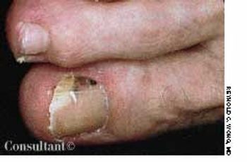
Transverse depressions like the one shown here appeared on all the other nails of this 68-year-old man several weeks after he had suffered a myocardial infarction.
Transverse depressions like the one shown here appeared on all the other nails of this 68-year-old man several weeks after he had suffered a myocardial infarction.
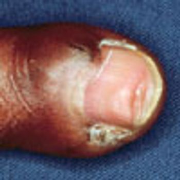
The left middle finger of this 30-year-old man was lacerated in a motorcycle accident. After it was surgically repaired, the finger developed some dystrophy as well as a small, separate fingernail in the lateral sulcus of the proximal nail fold.

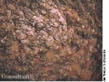
Eczema was the original diagnosis of this pink, scaly, asymptomatic patch on a 44-year-old man's chest. A mid-potency corticosteroid ointment had been prescribed but had no effect on the lesion. Now, 18 months later, the patient said the patch had been slowly enlarging.
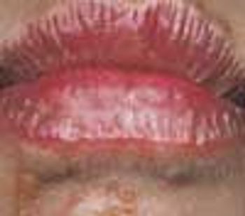
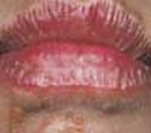
An 8-year-old boy was taken to his physician because of a rash around his mouth. After spending the day at the beach with his family, his lips had become red, itchy, and swollen. His sister had a similar, although milder, condition. Both children had been sucking on limes while at the beach. One week later, the boy experienced the chapped lips, patchy perioral erythema, swelling, and blisters.

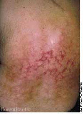
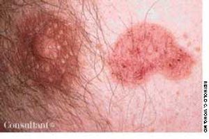
Consultation was sought for diagnosis of the concentric rings of erythema and scaling that appeared on one side of a 42-year-old woman's face. The patient had used a moderately potent topical corticosteroid that had been prescribed for a facial "rash."
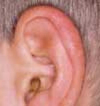
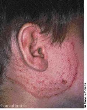
Each spring for the past 3 years, this boy has complained of an itchy ear rash. The dull red edematous papules develop on the outer portion of the helix, and the rash heals promptly after 2 weeks.
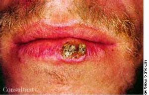
The tender, crusted nodule on the lower lip of a 35-year-old man had been growing slowly for 6 months. The patient reported a history of extensive sun exposure.
A 35-year-old man had a 5-year history of progressive hair loss characterized by follicular inflammation with destruction of the follicle and consequent permanent alopecia. Almost the entire scalp was involved. A few pustules were seen on examination, but the clinical picture was mostly one of scarring and irreversible hair loss.
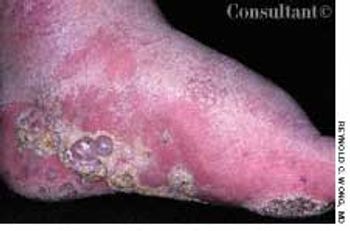
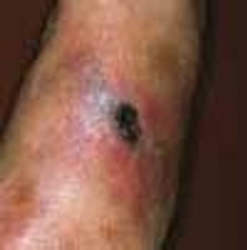
A 69-year-old man with a long history of lymphedema secondary to repeated cellulitis sought medical care for mildly pruritic, nontender, purple nodules that had erupted on the bottom and side of one foot 6 months earlier. Scale surrounded the nodules.

An otherwise healthy 34-year-old woman was concerned because of the abrupt onset of rapid hair loss, accompanied by scaling of the underlying skin. The disorder had begun 3 months earlier, and the right parietal and temporal areas were now red and swollen and had adherent scale. An antinuclear antigen titer was negative. Biopsy revealed changes consistent with lichen planus of the scalp, also known as lichen planopilaris and lichen planus follicularis.
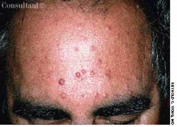
Asymptomatic facial and truncal papules began developing several years before this 55-year-old man sought medical care. The lesions were slightly yellowish or reddish, and many had a central punctum. Biopsy revealed a microscopic picture consistent with sebaceous adenoma.
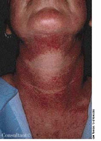
The coppery hyperpigmentation on the front and sides of a 47-year-old woman's neck, sparing a large area on the anterior surface, had been present for many years. She had no itching, burning, or other symptoms associated with the discoloration. Questioning revealed that she used perfume liberally on her neck and had been a sun worshipper in years past.
An area of mottled skin developed on the back of a 55-year-old woman who has had arthritis for several years. She often applied a heating pad to her middle and lower back for relief from arthritic pain.
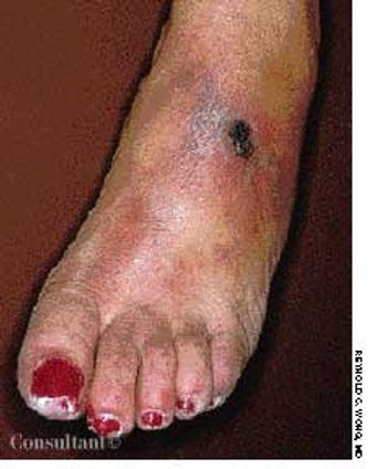
A patchy, macular, erythematous area developed on the dorsum of a 63-year-old woman's left foot 4 months ago. Because the lesion was asymptomatic and she was otherwise healthy, no workup was instituted at that time. The affected area subsequently became ulcerated, tender, and painful. The patient now had a 1.2-cm ulcer covered by a dried, hemorrhagic crust and surrounded by a livedo reticularis–like pattern. The entire area was extremely tender.
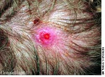
A red papule developed on the scalp of a 52-year-old man who had a history of adenocarcinoma of the lungs. The patient was a former cigarette smoker.
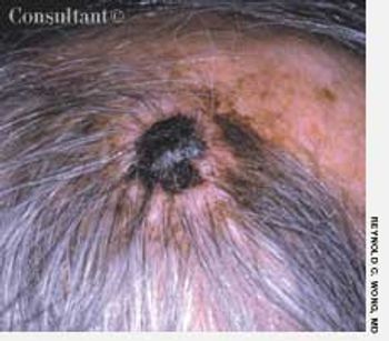
A previously asymptomatic, large black plaque on a 65-year-old man's scalp recently began to bleed. The lesion had grown considerably since it first appeared as a small black macule 3 years earlier.
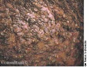
Brown-black discoloration and a soft, velvety papillomatosis of the axillary, inguinal, genital, and neck areas were seen in a 46-year-old man. He had hypertriglyceridemia, for which he had recently begun taking nicotinic acid. After a few days of therapy, he noticed the onset of this asymptomatic hyperpigmentation.
A 45-year-old woman who had had several melanomas removed was concerned because of hyperpigmentation beneath her toenail. Some distal onycholysis was also noted. Because of the patient's history and the fact that she could not definitely recall injuring the toe, a partial nail evulsion was performed. Fortunately, the only finding was dried blood.
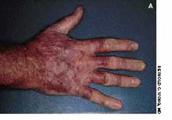
A 45-year-old man had a red, somewhat annular, slightly scaly plaque studded with red nodules that covered the back of his right hand. Nail dystrophy was evident on the middle finger. The patient's left hand was free of lesions, but the soles of both feet were slightly red and scaly, and there was an annular, serpiginous border on the left foot.
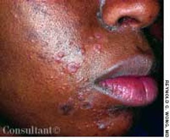
The widespread eruption of asymptomatic macules and flat, palpable, flesh-colored lesions prompted a 23-year-old woman to consult her physician. The lesions-some of which had dark centers-were concentrated on the patient's face, neck, and upper back; the palms, soles, anal mucosa, and genital areas were clear. The patient denied systemic symptoms. She was seronegative for HIV.
Cutaneous photosensitivity followed by acute, then chronic, skin lesions on sun-exposed areas characterizes this disorder.

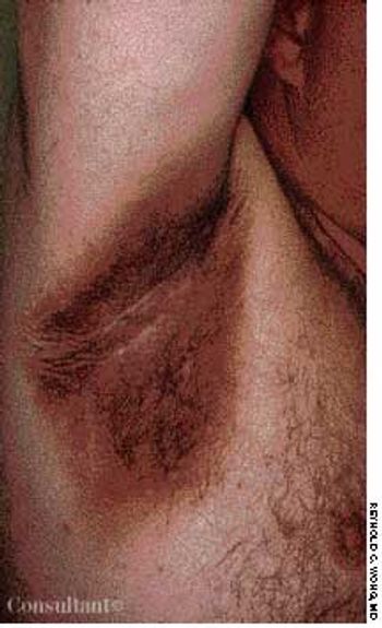
The dark red plaques seen here developed rapidly under both arms of this markedly obese 43-year-old woman. The lesions spread concentrically, forming necrotic ulcerations with overhanging borders, and there was surrounding violaceous discoloration. No other areas of the patient's body were involved, and she was otherwise in good health.
Four months after a patchy, macular, erythematous spot erupted on the dorsum of a 63-year-old woman's left foot, the area became ulcerated, tender, and painful. The 1.2-cm ulcer was covered by a hemorrhagic crust surrounded by a cyanotic reticular discoloration of the skin.
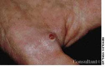
This red papule developed 6 months ago at the base of a 53-year-old woman's thumb. The lesion was asymptomatic, but it bled easily when traumatized. It was treated definitively with surgical excision, and the biopsy report was consistent with the clinical diagnosis of pyogenic granuloma. This most commonly appears on the face or fingers and may result from minor trauma.
Four months after a patchy, macular,erythematous spot erupted on thedorsum of a 63-year-old woman’s leftfoot, the area became ulcerated,tender, and painful. The 1.2-cm ulcerwas covered by a hemorrhagic crustsurrounded by a cyanotic reticulardiscoloration of the skin.
A previously asymptomatic, largeblack plaque on a 65-year-old man’sscalp recently began to bleed. The lesionhad grown considerably since itfirst appeared as
A 69-year-old man with a long history of lymphedema secondary to repeated cellulitis sought medical care for mildly pruritic, nontender, purple nodules that had erupted on the bottom and side of one foot 6 months earlier. Scale surrounded the nodules.
September 14th 2005
September 14th 2005
September 14th 2005

September 14th 2005
September 14th 2005
September 14th 2005