
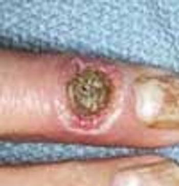
Cutaneous manifestations develop in approximately 30% of persons with diabetes. Premature atherosclerosis is a common complication of the disease that can cause peripheral infarction, ulceration, and necrosis.
Cutaneous manifestations develop in approximately 30% of persons with diabetes. Premature atherosclerosis is a common complication of the disease that can cause peripheral infarction, ulceration, and necrosis.

This rare condition affects both men and women. The average age at onset is 53 years. The lesions are deep brownish red to purple papules, nodules, and plaques. Blisters and ulcers also can occur.
A 39-year-old man who has sickle cell disease suffers with chronic ankle ulcers typical of the disorder. Ulcerations occur in approximately 50% of persons who are homozygous for sickle cell disease.
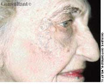
A 67-year-old woman sought medical treatment for the persistent, large “blackheads” on her face. Examination revealed diffusely thickened and yellowed skin with deep furrows. The periorbital and malar areas were studded with large open comedones.
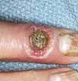
A 16-year-old boy removed a small sliver of wood from the palm of his hand with a pocket knife. However, the fledgling “surgeon” created a small puncture wound during this operation. Over the next month, a small, moist, friable papule grew at the site.
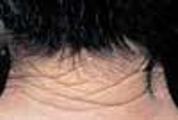
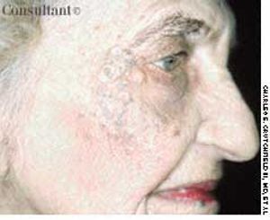
During a routine skin cancer screening, yellowed, thickened, leathery skin was noted on the posterior neck of a 73-year-old retired construction worker. Colloquially, this condition is called “sailor's skin” or “farmer's skin” and is seen in persons who have had long-term exposure to the sun. It is known clinically as cutis rhomboidalis nuchae, because the well-defined furrows in the skin resemble an irregular rhomboidal pattern.
A 41-year-old man sought treatment of the rough, intensely itchy patch on the dorsum of his left foot and lateral ankle. There were no other similar plaques on his body.
A 17-year-old high school athlete was anxious about this new “mole” that appeared on the heel of his right foot. His concern was prompted by the recent diagnosis of melanoma in his aunt.
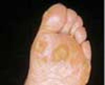
A 63-year-old farmer first noticed multiple rough bumps on his hands and feet at least 20 years before pointing them out to his physician. A diagnosis of arsenical keratoses was made after the patient reported that as a child he had worked on his family's potato farm, where a commonly used pesticide, “Paris Green,” was applied to the plants. The active ingredient in this pesticide was inorganic arsenic.
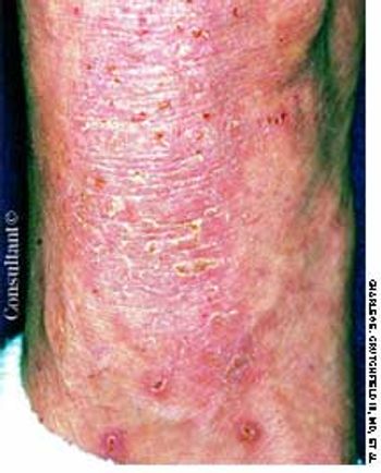
A 40-year-old farmer had been complaining for 3 weeks of a tender, red, itchy, scaling plaque with papulopustules on one knee. A potassium hydroxide examination of the scale revealed fungal hyphae.
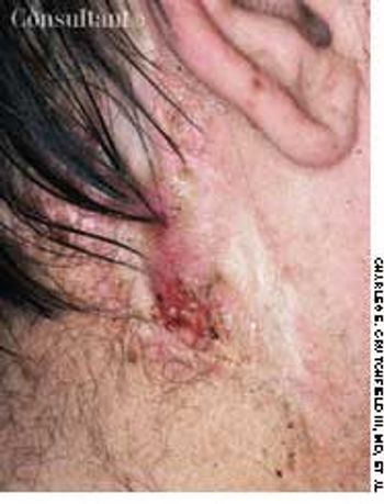
An ulcerating nodule on his left pre-auricular region worried a 64-year-old man.
A scaling, red, fissured area between digits 4 and 5 on her right hand sent a 33-year-old woman to her physician. Diagnosis of interdigital Candida was confirmed by a potassium hydroxide evaluation of material from the site.
This 19-month-old child exhibits the characteristic features of hypomelanosis of Ito, a neurocutaneous disorder.
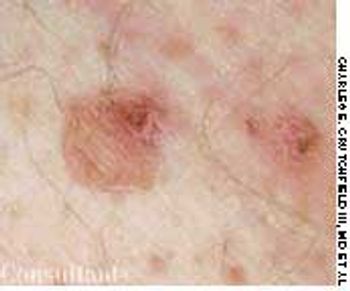
A 48-year-old man requested medical evaluation of the multiple spots that gradually appeared on his legs over the past 5 years.
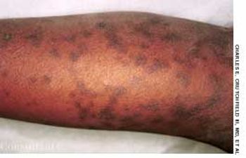
Flat, tan-pink patches on his lower legs disturbed a 52-year-old man. The lesions had visible, nonpalpable petechiae, which did not blanch on diascopy; telangiectasia and inflammatory vasodilation, therefore, were excluded from consideration.
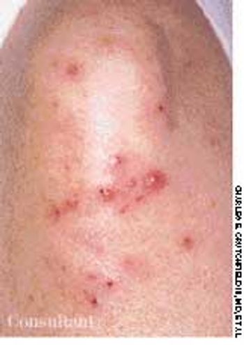
Flesh-colored to red-brown nonpruritic papules developed most prominently on the elbows, forearms, and knees of a 2 1⁄2-year-old boy. The rash was preceded by a low-grade fever and mild, upper respiratory tract symptoms.

A 23-year-old man complained of unsightly “flat warts” all over his chest. These lesions first appeared in early adolescence. The clinical suspicion of epidermodysplasia verruciformis was confirmed by a biopsy.

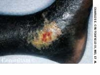
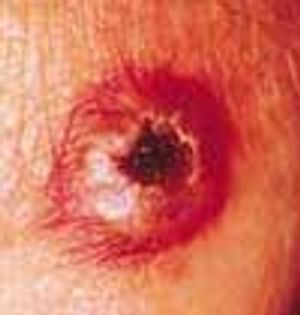
A 61-year-old man with a history of squamous cell carcinoma was concerned about a tender nodule on his ear. He complained of exquisite tenderness with pressure, such as when lying on the affected side at night.
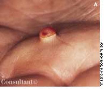
Concerned about this unsightly and painful lesion, a 34-year-old man asked to have it removed. The patient first noted this eruption on his back several months before. This lesion is an infundibular cyst.
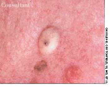
A 57-year-old man with a history of venous stasis leg ulceration wondered about the “white spots” on his leg.
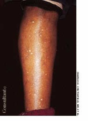
After more than 15 years of wondering what the “white specks” on his legs were, a 64-year-old man consulted his physician. The patient was taking medication to control hypertension; he was otherwise healthy.
A 57-year-old man with a history of venous stasis leg ulcerationwondered about the “white spots” on his leg. Thecondition is atrophie blanche, which manifests as smooth,ivory-white macules and plaques of sclerosis stippled withtelangiectasia that often are surrounded by mild to moderatepigmentation.
Cutaneous manifestations develop inapproximately 30% of persons withdiabetes. Premature atherosclerosisis a common complication of thedisease and can cause peripheral infarction,ulceration, and necrosis.
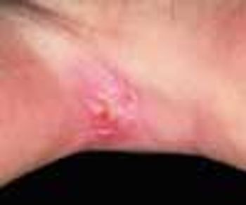
A hyperpigmented macule on the submental surface disturbed this 56-year-old man.
A 54-year-old Asian woman complained of itching from a newly erupted rash.
September 14th 2005
August 1st 1999
September 14th 2005

September 14th 2005
September 14th 2005
August 1st 1999