


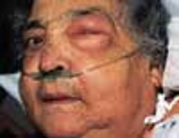
A 68-year-old woman was admitted to the hospital with rapidly increasing, painful swelling of the left eye. She had moderately severe, corticosteroid-dependent chronic obstructive pulmonary disease.
A 68-year-old woman was admitted to the hospital with rapidly increasing, painful swelling of the left eye. She had moderately severe, corticosteroid-dependent chronic obstructive pulmonary disease.
For 2 months, a 22-year-old uncircumcised man noticed an asymptomatic, erythematous, static lesion on the glans penis. He had applied an over-the-counter “jock-itch” ointment for 2 weeks but to no avail. The young man was otherwise healthy and denied having dysuria or a history of sexually transmitted disease.
A 24-year-old man presented for evaluation of pruritic vesicles on both feet. Ten days earlier, dyshidrotic eczema had been diagnosed by another physician who prescribed triamcinolone ointment. The patient reported that the foot eruption worsened after the topical medication was applied.
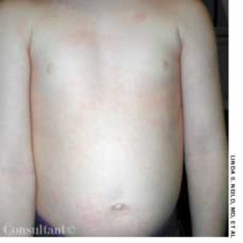
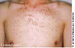
This 6-year-old boy was brought to his physician for evaluation of a rash. The child had been running a fever and, for the past 48 hours, had been complaining of a sore throat, headache, and abdominal pain.
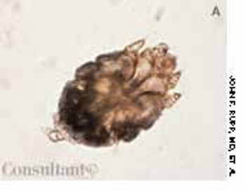
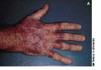
Persistent, unremitting itch-which intensifies at night-is the chief complaint of patients with scabies. The female Sarcoptes scabiei mite (A) burrows into the stratum corneum, where she lays eggs. The parasite is transferred by intimate contact and fomites, such as infested clothing, towels, and bedding.
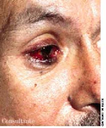
A painful swelling over the right lower eyelid with conjunctival injection was evaluated in a 28-year-old injection drug user. Cephalexin and corticosteroid ophthalmic drops failed to resolve the condition.
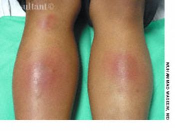
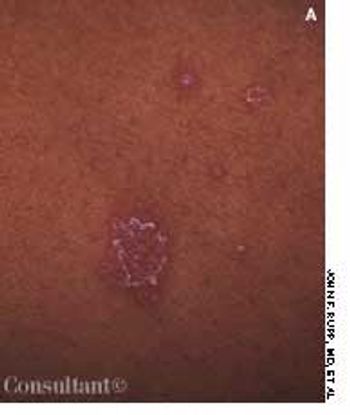
A 16-year-old girl had had tender, erythematous, nodular, shiny lesions on the extensor aspect of both shins for 2 weeks. There were no ulcerations or adenopathy. She denied fever, cough, sore throat, pruritus, and GI symptoms. Aside from oral contraceptives, she was not taking any medications.

A comatose 29-year-old woman was brought to the emergency department. Her family reported that she had been well until 4 days earlier, when headache and fever developed. She went to another hospital at that time and was told she had an abscessed tooth. She was given erythromycin, and the tooth was extracted the following day. The patient's headache and fever worsened; a sore throat also developed, and a rash appeared on her trunk, arms, and legs. The family denied any HIV risk factors, unusual medical history, recent travel, and exposure to persons with infectious diseases.
This self-limited eruption is characterized by erythematous, scaling, oval-shaped macules on the trunk and proximal extremities. Most outbreaks begin with a single, large patch-a mother or herald patch-that typically is found on the trunk. Commonly, this lesion is confused with ringworm.
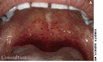
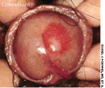
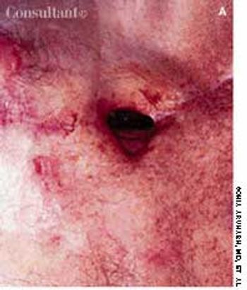
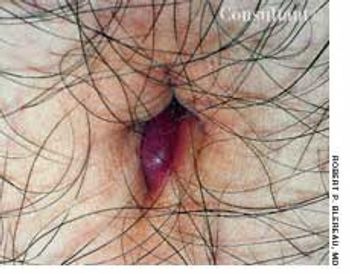
The variable appearance of palatal petechiae is demonstrated by these two cases, presented by Robert P. Blereau, MD of Morgan City, La. The petechiae appear as discrete pinhead lesions in a 10-year-old boy, whereas they are manifested as minute hemorrhagic areas in a 30-year-old woman.

While playing outside, a 23-month-old girl became sleepy and difficult to arouse. The mother brought her daughter to the emergency department (ED); posturing and a dilated and fixed right pupil were noted. The child was hospitalized.
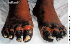
The left hand of a 45-year-old man was affected with the characteristic intact bullae and dried, crusted, ruptured bullae of bullous impetigo.
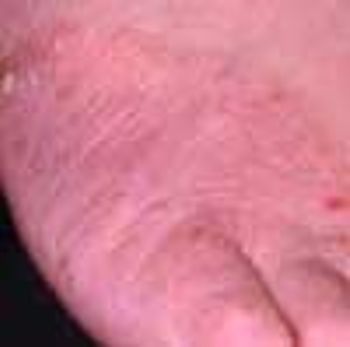
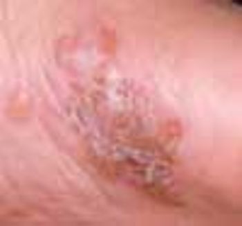
A pruritic rash of unknown origin had been present on the dorsum of a 26-year-old woman's foot for several months. Despite oral antibiotic therapy and applications of antifungal creams and topical corticosteroids, the condition did not resolve.

Congenital adrenal hyperplasia is an autosomal recessive disorder. Deficiency of 21-hydroxylase accounts for 95% of all cases.
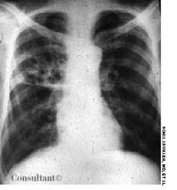
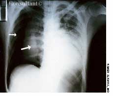
Having been treated for pulmonary tuberculosis (TB) 25 years earlier, a 60-year-old man (a nonsmoker) now complained of a chronic cough. The cough was occasionally accompanied by yellowish sputum but no hemoptysis. Examination revealed persistent coarse crackles in the right posterior hemithorax, and the x-ray study seen here established the diagnosis of cystic bronchiectasis.
A 45-year-old man had a red, somewhat annular, slightly scaly plaque studded with red nodules that covered the back of his right hand. Nail dystrophy was evident on the middle finger. The patient's left hand was free of lesions, but the soles of both feet were slightly red and scaly, and there was an annular, serpiginous border on the left foot.
For the past 2 days, a 30-year-old man had experienced scant hemoptysis. He had also lost a significant amount of weight-5 kg (11 lb)-over the last 2 months. The patient, a recent emigrant from Ecuador, had no history of tuberculosis (TB) or of exposure to this disease, and he had not been skin tested with purified protein derivative (PPD). He denied exposure to risk factors for HIV infection.
An 82-year-old man underwent right pneumonectomy for squamous cell carcinoma of the right lower lobe. His postoperative course was complicated by prolonged air leak from the chest tube, suggesting a bronchopleural fistula secondary to leakage from the bronchial stump. Over the ensuing 3 months, the air leak slowly decreased, but the patient was left with a nonhealing scar on the anterior thoracic cavity, as seen here.
An obese 17-year-old boy sought treatment of an apparent abscess of the umbilicus. Hot soaks, black salve, and oral cephalexin were prescribed. Although there was some drainage, the lesion persisted and the patient returned for further evaluation.
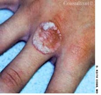
For 3 weeks, a 14-year-old boy had been aware of an enlarging lesion on the back of his hand. He recalled no trauma to the affected area. Further questioning by Dr D. Keith Cobb of Savannah, Ga, revealed that a 4-mm verruca, or wart, had been removed from the same site 6 months earlier with cryosurgery by a different physician.
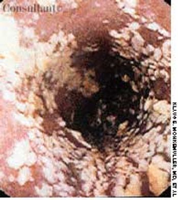
An obese 52-year-old woman with a 5-year history of type II diabetes mellitus had odynophagia and dysphagia for several days. She described the sensation as food “sticking” in her chest. She also complained of vaginal itching, polyuria, and polydipsia. The only remarkable finding on physical examination was candidal vaginitis. The patient did not smoke cigarettes or drink alcoholic beverages, and there was no history of recent weight loss.
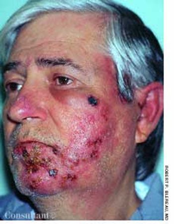
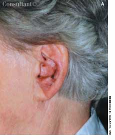
Redness and swelling of the left cheek, chin, and ear bothered a 51-year-old man. The initial diagnosis was cellulitis and/or allergic dermatitis; oral amoxicillin/clavulanate, 500 mg tid, and a low- to medium-potency corticosteroid cream, alclometasone, were prescribed. Within 1 to 2 days, pimples emerged in the reddened areas and rapidly crusted.

A 4-year-old girl is brought to the emergency department with a pruritic rash of 24 hours' duration. Her mother reports that the lesions developed after the child ate strawberries.

A mother, fearing that her 4-year-old son had been abused at his day-care center, rushed him to the emergency department, where an evaluation revealed a platelet count of 1,000/µL. Except for bruises on the boy's face and legs, the physical findings were normal. Bone marrow aspiration showed numerous megakaryocytes and was otherwise normal. The youngster's history included treatment for bronchitis, sinusitis, and conjunctivitis 2 weeks earlier.

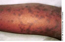
For several weeks, a 78-year-old woman had an intensely pruritic, diffuse, raised, slightly scaly, erythematous rash that persisted despite the use of several over-the-counter topical medications (such as hydrocortisone and clotrimazole cream). Since her last visit about 3 months earlier for a blood pressure reading, she had been well except for 2 episodes of night sweats. For several years, she had been taking levothyroxine and reserpine/hydrochlorothiazide; about 6 months ago, valsartan/hydrochlorothiazide had been prescribed.