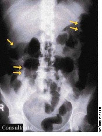
Infectious Disease

Latest News


CME Content


For 3 weeks, a 14-year-old boy had been aware of an enlarging lesion on the back of his hand. He recalled no trauma to the affected area. Further questioning by Dr D. Keith Cobb of Savannah, Ga, revealed that a 4-mm verruca, or wart, had been removed from the same site 6 months earlier with cryosurgery by a different physician.

An obese 52-year-old woman with a 5-year history of type II diabetes mellitus had odynophagia and dysphagia for several days. She described the sensation as food “sticking” in her chest. She also complained of vaginal itching, polyuria, and polydipsia. The only remarkable finding on physical examination was candidal vaginitis. The patient did not smoke cigarettes or drink alcoholic beverages, and there was no history of recent weight loss.

Redness and swelling of the left cheek, chin, and ear bothered a 51-year-old man. The initial diagnosis was cellulitis and/or allergic dermatitis; oral amoxicillin/clavulanate, 500 mg tid, and a low- to medium-potency corticosteroid cream, alclometasone, were prescribed. Within 1 to 2 days, pimples emerged in the reddened areas and rapidly crusted.

Urticaria

A 4-year-old girl is brought to the emergency department with a pruritic rash of 24 hours' duration. Her mother reports that the lesions developed after the child ate strawberries.

A mother, fearing that her 4-year-old son had been abused at his day-care center, rushed him to the emergency department, where an evaluation revealed a platelet count of 1,000/µL. Except for bruises on the boy's face and legs, the physical findings were normal. Bone marrow aspiration showed numerous megakaryocytes and was otherwise normal. The youngster's history included treatment for bronchitis, sinusitis, and conjunctivitis 2 weeks earlier.

For several weeks, a 78-year-old woman had an intensely pruritic, diffuse, raised, slightly scaly, erythematous rash that persisted despite the use of several over-the-counter topical medications (such as hydrocortisone and clotrimazole cream). Since her last visit about 3 months earlier for a blood pressure reading, she had been well except for 2 episodes of night sweats. For several years, she had been taking levothyroxine and reserpine/hydrochlorothiazide; about 6 months ago, valsartan/hydrochlorothiazide had been prescribed.

A mildly painful, nonpruritic rash on the forearms and legs prompted a 42-year-old man to go to the emergency department (ED). The patient noted the rash when he awoke that morning. He had had joint pain and fever for the past 7 days and generalized malaise with chills that began about 3 days earlier. He had no significant medical history.

A previously healthy 51-year-old man presented with weight loss and poor appetite of 2 months' duration. He was heterosexual and had many sexual partners. Except for a temperature of 38.3°C (100.9°F) and left basal rhonchi, results of physical examination were normal. A chest radiograph and CT scan, as seen here, showed large cavitary lesions in the lower left lobe.

A 40-year-old man was hospitalized with the superficial, reddening, and painful facial lesion seen here. Its borders were sharp, and it had developed rapidly. The patient had a temperature of 40°C (104°F) and chills. His erythrocyte sedimentation rate was elevated, and he had neutrophilic leukocytosis.

A 40-year-old man, originally an Ohio resident, underwent a chest x-ray study during medical clearance following surgery to repair a hernia. Although the patient had no pulmonary symptoms, the film showed multiple small, punctate calcifications in both lung fields.

After 1 week of scratching a pruritic, reddish rash on a swollen hand and enduring a “burning sensation,” a 43-year-old man visited his physician. The patient worked as a meatpacker. He had no other medical conditions, took no medications, and had no known drug allergies.

Pain, swelling, and purulent, blood-stained drainage around the lower back had plagued a 21-year-old woman for 3 weeks. On examination, multiple openings were seen overlying the natal cleft, the sacrococcygeal region, and the upper part of the midline. The last was the area of drainage. Insertion of a metal probe at that point revealed a pilonidal sinus that communicated with centrally located openings.

A 32-year-old Hispanic woman with AIDS presented with a 1-month history of diarrhea; abdominal bloating and cramps; loss of appetite; and pronounced fatigue, malaise, and weight loss. She had no fever or chills and was not vomiting. Her CD4+ cell count was 12/µL. Results of a routine microscopic examination of stool for ova and parasites were negative; an acid-fast stain of stool demonstrated oocytes of Cyclospora cayetanensis measuring 8.8 mm in diameter (pictured, magnification ×1,000). This is about twice the size of the Cryptosporidium parvum oocyte, which typically is 4 to 5 mm.

Asteatosis

Erythematous, scaly lesions with double-edged borders had been present on a 14-year-old boy's left upper arm and lower legs for about a year. The lesions were occasionally pruritic, and some resembled ringworm. At times, fine yellow crusting suggestive of impetigo was present. The boy took very hot baths and showers.

Fever occurring twice daily, accompanied by profuse sweating, anorexia, and malaise of 15 days' duration resulted in hospitalization of a 31-year-old woman. At this time, her temperature was 37.6°C (99.7°F); blood pressure, 110/70 mm Hg; pulse rate, 90 beats per minute; and respiratory rate, 18 breaths per minute. A palpable spleen was about 3 to 5 cm below the left subcostal margin. Neither lymphadenopathy nor hepatomegaly was noted.

Tinea Faciei

An eruption on the face of a 49-year-old woman had been misdiagnosed as a staphylococcal infection; the rash failed to respond to oral and topical antibiotics. A mid-potency topical corticosteroid also had been tried, but the eruption worsened.

The widespread eruption of asymptomatic macules and flat, palpable, flesh-colored lesions prompted a 23-year-old woman to consult her physician. The lesions-some of which had dark centers-were concentrated on the patient's face, neck, and upper back; the palms, soles, anal mucosa, and genital areas were clear. The patient denied systemic symptoms. She was seronegative for HIV.