


The parents of a 16-year-old girl report that during the past several months, she has exhibited behavioral changes, irritability, increased anger, depression, and anxiety. The girl had previously been healthy, and there has been no recent illness or trauma.
Infectious Disease
Latest News
Advertisement
Advertisement
HANDY LIST TO KEEP
A tender nodule; an itchy truncal rash; multiple pruritic eruptions--can you identify the disorders pictured here?
ABSTRACT: Early treatment with disease-modifying anti-rheumatic drugs (DMARDs)--alone or in combination-- can prevent joint damage and minimize disability. Until recently, the DMARDs used predominantly in patients with rheumatoid arthritis had been methotrexate, sulfasalazine, and hydoxychloroquine. Older DMARDs such as gold, d-penicillamine, and azathioprine have fallen out of favor because of their long- term toxicities or modest benefit. Six newer DMARDs--leflunomide, etanercept, infliximab, adalimumab, rituximab, and anakinra--have greatly expanded the current treatment options.
Abstract: All children with asthma should have periodic office visits, usually every 3 to 6 months, in which asthma action plans are updated. Periodic assessment of lung function by peak expiratory flow or office spirometry can help determine the appropriate treatment strategy. Low daily doses of inhaled corticosteroids remain the first and most effective choice of therapy for persistent asthma. If this approach is inadequate, adding a second medication, such as a leukotriene modifier or a long-acting ß2-agonist, is suggested. Short-acting ß2-agonists remain the most important therapy for intermittent asthma. For most children, the best route is via a metered-dose inhaler with either a spacer or valved holding chamber. If these agents are inadequate, a short course of oral corticosteroids may be required. (J Respir Dis. 2005;26(8):348-358)
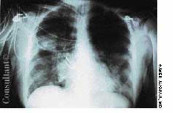
32-year-old man presents with a 4-day history of fever (temperature as high as 38.8 C to 39.4 C with severe rigors, chills, and profuse night sweats; generalized myalgias, including dull, aching headache; and dry cough.
A 54-year-old woman with a history of hypertension presented with a worsening headache and a left hemisensory defect. A CT scan of her head without contrast showed a right parietal hemorrhage with spreading edema; the masslike effect caused shifting of the midline to the contralateral side. The patient gradually became comatose and required intubation for airway protection. Intravenous corticosteroids were administered to decrease the effect of the lobar hemorrhage. Fever developed 3 days after admission.
A 79-year-old man has an elevated prostate-specific antigen (PSA) level(11.3 ng/mL). About 1 month earlier, when he was hospitalized for a seriousurinary tract infection (UTI), his PSA level was 13.3 ng/mL. The more recentlevel was obtained after he received antibiotic therapy for the UTI.
Abstract: The laryngeal mask airway (LMA) and intubating LMA are valuable alternatives in patients in whom intubation has failed and who need oxygenation and ventilation immediately. The dual-lumen, dual-cuffed airway tube is effective in a variety of settings and can tolerate ventilation at pressures as high as 50 cm H2O; it is contraindicated in awake patients who have intact airway reflexes, caustic ingestions, and upper airway obstruction from a foreign body or pathology. Surgical airways are lifesaving techniques when intubation is unsuccessful or impossible through the mouth or nose. It may be particularly appropriate in patients with laryngeal or facial trauma, upper airway obstruction, or oropharyngeal injury. When patients aged 12 years and older cannot be ventilated by mask or intubated with traditional methods, surgical or needle cricothyrotomy is the procedure of choice. (J Respir Dis. 2005;26(7):298-302)
Inflammation plays a major role in coronary artery disease (CAD), whereby inflammatory changes develop in the blood vessel walls.1 This observation has spurred interest in exploring the connection between CAD and markers of inflammation, including C-reactive protein (CRP), fibrinogen, serum amyloid A, and many other novel markers.
The authors describe a rare cause of diffuse thoracic lymphadenopathy--Cogan syndrome. This case was remarkable for the temporal development of extensive lymphadenopathy independent of other hallmark symptoms and signs of this syndrome. In the appropriate clinical setting, Cogan syndrome should be considered in the differential diagnosis of thoracic lymphadenopathy.
Abstract: Although smoking cessation is still the most impor- tant intervention in chronic obstructive pulmonary disease (COPD), a variety of pharmacologic therapies are available to help manage symptoms. Short-acting ß2-agonists and/or ipratropium should be taken as needed, and the use of additional therapies is based on the severity of disease. Patients with moderate or severe COPD should regularly take 1 or more long-acting bronchodilators. The long-acting ß2-agonists salmeterol and formoterol have been demonstrated to improve health-related quality of life. Newer therapies include the long-acting anticholinergic tiotropium and a salmeterol-fluticasone combination. These agents improve forced expiratory volume in 1 second and may reduce the rate of acute exacerbations. For patients with moderate to very severe COPD, participation in a pulmonary rehabilitation program can improve health status, quality of life, and exercise tolerance. (J Respir Dis. 2005;26(7):284-289)
A 39-year-old African American man complains of mild dyspnea that has worsened steadily over the past 2 weeks and that is unrelated to activities or unusual exposures. He denies trauma, fever, and chest pain. His medical history is unremarkable.
Stories about community-associated infections with methicillin-resistant Staphylococcus aureus (MRSA) have been making headlines in recent months in both the medical and popular press. A familiar problem in hospitals and nursing homes for decades, growing numbers of MRSA infections have been documented over the past few years in prison inmates, soldiers, athletes, Pacific Islanders, Alaska Natives, Native Americans, and men who have sex with men.
Multiple sclerosis, the most common cause of disability among young adults, can take years to diagnose because symptoms may wax and wane over time, and patients may not initially seek evaluation.
9-year-old girl was seen in the emergency department (ED) with a 5-day history of non-bilious, non-projectile emesis, decreased appetite, and persistent right lower quadrant pain following an appendectomy 5 days earlier.
A 42-year-old woman presents with severe diarrhea that began 3 days earlier and has become progressively more severe. She is now having 10 or more watery bowel movements per day. She has had moderate nausea but no emesis, hematemesis, or hematochezia.
ABSTRACT: The management of chronic daily headache is difficult and complex. Those affected have a sensitive nervous system, and their predisposition for a low tolerance to sensory stimuli appears to be inherited. Under appropriate conditions, the equilibrium or balance between bombardment from painful stimuli and the regulatory systems that inhibit those stimuli is disrupted, allowing painful stimuli to become manifest at a greater intensity than in the nonmigraineur. Successful management depends on close adherence to nonpharmacologic approaches and pharmacologic regimens that desensitize the system and restore equilibrium. Comorbid conditions must be identified and treated as well.
To help lubricate the epithelial surface of the nose, mucus must move steadily toward the pharynx, where it is eventually swallowed. If it stagnates in the nasal or sinus cavities, it can dry out or become infected.
In his Photo Essay case of a 5-year-old girl with a herpes simplex virus (HSV) infection on her thumb , Dr Robert Blereau writes that treatment consisted of mupirocin ointment. Frankly, I was surprised to read that this agent was used to treat an HSV infection. Was this the only treatment?
A 5-year-old girl is brought for evaluation of an asymptomatic inflamed streak on one leg that has been present for several weeks. She is otherwise healthy and takes no medications.
Children are at greater risk than adults for many travel-related problems, such as barotitis and barotrauma associated with flying, cold and heat injury, drowning, and infection with geohelminths. Most of these problems can be avoided with appropriate measures. Here, a summary of the most important steps.
The American Thoracic Society (ATS) and the Infectious Diseases Society of America recently published guidelines for the management of hospital-acquired pneumonia (HAP).1 These guidelines, which are an update of a 1996 ATS consensus statement,2 focus on bacterial HAP in immunocompetent adults. This includes ventilator-associated pneumonia (VAP) and health care-associated pneumonia (HCAP). Selected highlights are presented here.
Anorectal abscesses and fistulae, pilonidal disease, rectal prolapse, pruritus ani, and anal masses are discussed, with an emphasis on diagnosis and treatment of these conditions in the primary care office setting.
Anorectal complaints are commonly seen in primary care practice. The two most common anorectal conditions are hemorrhoids and anal fissures.
Advertisement
Advertisement
Trending on Patient Care Online
1
Newer GLP-1 Therapies Linked to Fewer Alcohol-Related Hospitalizations in Adults With AUD
2
2026 Dyslipidemia Guideline Expands Statin Eligibility to 21.5 Million More US Adults
3
Early Liver Disease Detection Starts in Primary Care: A Q&A With Stevan Gonzalez, MD
4
Compulsive Smartphone Use Linked to Depressive Symptoms in Older Adults
5