


An 85-year-old man was admitted to the hospital with acough and shortness of breath of 1 week’s duration anda fever and increased sputum production for 2 days. Hishistory included renal cell carcinoma and metastatic renalcancer for 2 years. The patient had smoked cigarettesfor 30 years. He had lost 30 lb during the last few months.A chest film revealed pneumonia of the right lowerlobe. Metastatic nodules were noted on the scalp; extensivelung, bone, and brain metastases also were found.
Dermatology
Latest News
Advertisement
Advertisement
A 40-year-old man was concerned about an enlarging painlessmass on the right side of his neck that had been presentfor 6 months (A). The patient reported no other healthproblems; his medical history was unremarkable, and hewas taking no medications.There was no family or personal history of thyroiddisease or of exposure to radiation. Thyroid function testresults were within normal limits. A chest film revealed nopathology.
Signs and symptoms that strongly suggest peripheral arterial occlusive disease include diminished or absent pedal pulses, a unilaterally cool limb, and atrophic skin that is shiny and hairless. An ankle-brachial index of less than 0.5 suggests multisegment disease. Management goals are to decrease functional impairment, treat underlying atherosclerosis, and control risk factors. Smoking cessation is imperative. A graduated walking program is a mainstay of treatment and is associated with greater improvement in pain-free walking than is drug therapy. Surgery and percutaneous intervention are generally reserved for patients with lifestyle-limiting claudication, ischemic pain at rest, tissue loss, or gangrene.

Several times a year a rash erupts on the chest, axillae, and neck of a 41-year-old woman. Her father and siblings have a similar history. A biopsy of the affected skin confirmed the suspected diagnosis of benign familial pemphigus, which is also called Hailey-Hailey disease.

Diffuse petechiae suddenly arose on the back and abdomen of a 79-year-old woman. Within several days, the asymptomatic lesions covered her arms and face as well.
A 40-year-old woman who said she had asthma was admitted to the hospital with worsening dyspnea and cough. A β-adrenergic agent was her only medication. The patient denied cigarette smoking and alcohol consumption. Except for an appendectomy 20 years earlier, her medical history was unremarkable. The patient was afebrile.

A 79-year-old man was brought to the emergency department with mental status changes. A CT scan revealed a lacunar infarct. Numerous asymptomatic nodules had been present for many years on the patient’s scalp and forehead.
For 2 years, a slightly pruritic, light brown, scaly rash had been present on a 20-year-old man’s neck. During the past 8 months, the eruption spread to the upper chest and upper arms. The patient reported that the rash changes color with the seasons. Multiple round to oval, hypopigmented, slightly scaly macules were noted on the neck, chest, and upper arms. Tinea versicolor was strongly suspected.

For 3 years, a linear plaque had been slowly developing on the left palm of a 47-year-old woman who had difficulty in opening and closing her hand. There was no contributory family history.
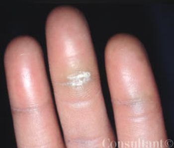
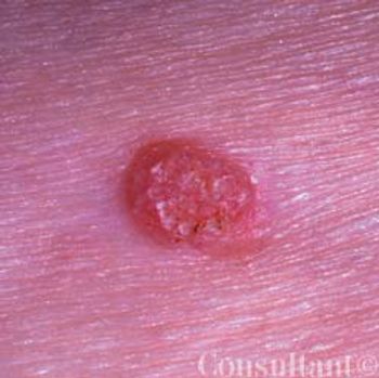
The 2-mm, white, very slightly raised lesion on the vermilion of a 37-year-oldman’s lower lip had been present for 2 months. The lesion was asymptomatic.There was no history of injury or burn to the area.

An uncircumcised 58-yearold man presented with a persistent “rash” on his penis of 5 years’ duration. He complained of localized irritation with coitus. Over-the-counter ointments and corticosteroid preparations had failed to clear the eruption. The patient had hypertension, hyperlipidemia, and coronary artery disease. He had been monogamous for the last decade and denied any risk factors for sexually transmitted diseases.
A 34-year-old woman (gravida 3, para 2) presented at 28 weeks’ gestation with a 3-week history of a pruritic rash that had progressively worsened. Multiple vesicles and bullae were noted; erosions and crusts on older lesions were also present. The patient had had no prodromal symptoms; she denied fever, chills, nausea, and vomiting. The purplish hue on her body resulted from application of the topical antibacterial agent, gentian violet, which the patient had obtained in Mexico.
A 38-year-old man was concerned that the small lesions on his lips were flatwarts. A biopsy identified Fordyce, or sebaceous, glands of the lips.
The soft “bubble” on the mucosal surface of a 42-year-oldman’s lower lip had developed, disappeared for 3 months,and returned. The lesion caused no pain or discomfort.
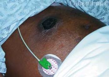
A 78-year-old man with a history of asthma, coronary artery disease, chronic obstructive pulmonary disease (COPD), and recently diagnosed prostate cancer was admitted to the medical ICU with exacerbation of COPD. The obtunded patient was unable to provide a history. A 4 × 6-cm firm nodule of unknown duration was noted over the right upper quadrant of the patient’s abdomen. There were no other cutaneous lesions.
A 38-year-old African American man presented with a 10-year history of a solitary �“wart” that had not responded to over-the-counter acid treatments. No other lesions were noted on the patient. Based on the clinical appearance, punctate porokeratosis was diagnosed.
A 1-year history of “dents” on her body prompted a 40-year-old African American woman to seek medical evaluation. Her medical history was unremarkable; however, there was a strong family history of severe type 2 diabetes mellitus. The patient denied having received or having self-administered injections into the affected areas. She reported no history of deep, tender nodules at the sites.
Intense pruritus centered around a sparse “rash” sent a 32-year-old Chinese man for medical evaluation. The patient’s medical and social histories were unremarkable.

A 62-year-old obese woman with adult-onset type 1 diabetes mellitus had a 6-year history of bilateral leg edema. During the last year, the edema worsened and the skin on her legs thickened. She also had multiple, bilateral, painful, superficial ulcers that drained copiously.
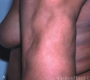
A 12-year-old African American girl comes to youroffice for a well-child checkup. She is tall for herage (height above the 95th percentile) and obese(body mass index [BMI] above the 95th percentile).Physical examination reveals acanthosisnigricans on her neck, axilla, and upper abdominalregion (Figure) and a vaginal yeast infection.Routine urinalysis reveals a glucose level ofgreater than 1000 mg/dL, with negative proteinand ketones. A random blood glucose test, obtainedbecause of the glucosuria, is 249 mg/dL.
A slightly pruritic, red, scaly rash on an 8-year-old boy’shands has been progressively worsening since it appeared4 months earlier. Nail pitting also was noted. There are noother rashes on his body. The patient is active in sports;denies any new exposure to soaps, clothing, or other contactants;and spends time in the homes of his recently divorcedparents.
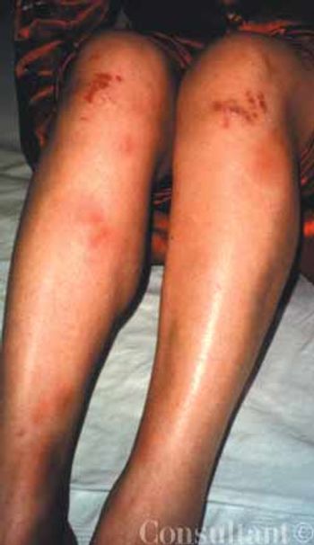
This condition features acutely tender nodules, marked erythema, and contusions that appear as a consequence of inflammation of subcutaneous fat.
This asymptomatic lesion had been present on a 75-year-old man's right buttock for 2 to 3 years. The 0.5 cm in diameter nodule featured an irregular, flesh-colored surface.
Reiter Syndrome (also called reactive arthritis) manifests as peripheral arthritis that is sometimes accompanied by such extra-articular findings as urethritis, conjunctivitis, and uveitis
Psoriatic Arthritis develops in approximately 20% of patients who have psoriasis.
Advertisement
Advertisement
Trending on Patient Care Online
1
Reishi Mushroom Extract Outperformed Melatonin for Chronic Insomnia in New Study
2
GLP-1 Insurance Barriers Are Reshaping OSA Diagnostic Referral Patterns
3
Interpreting Smartwatch Sleep Metrics in Menopausal Patients, With Fiona Baker, PhD
4
Home Sleep Testing for GLP-1 Coverage Shows Modest Diagnostic Yield in New Study
5