


When patients complain of malodoroushidrosis of the feet and they havetried every over-the-counter remedy,suggest they apply a regular underarmdeodorant/antiperspirant to theirfeet after showering
Dermatology
Latest News
Advertisement
Advertisement
A 67-year-old woman with insulin-dependent diabetesmellitus and uncontrolled hyperglycemia complained offatigue and malaise. For 2 years, a draining ulcer hadbeen present on the bottom of her left foot.
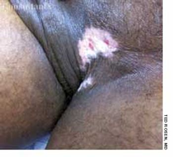
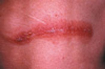
A 46-year-old man complained of “irritation” in the groin of several months’ duration. Ted Rosen, MD, of Houston noted a tender, macerated, hypopigmented plaque at the junction of the scrotum and upper inner thigh. At the periphery of the lesion was some detectable erythema and within the plaque were several small, superficial erosions.
A 20-year-old woman presents with a 3-week history of a pruritic, progressivelyenlarging erythematous lesion on one arm. She has a cat and recentlystarted horseback riding lessons. She is otherwise healthy and takes nomedication.
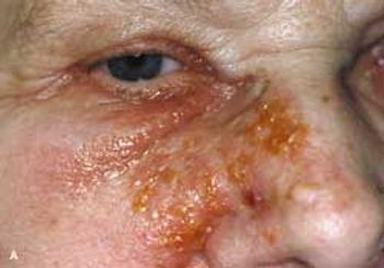
For a few days, this 73-year-old woman had had an itchy, painful rash on the right side of her face. Despite its proximity to her eye, she had no ocular involvement and no blurring of vision.
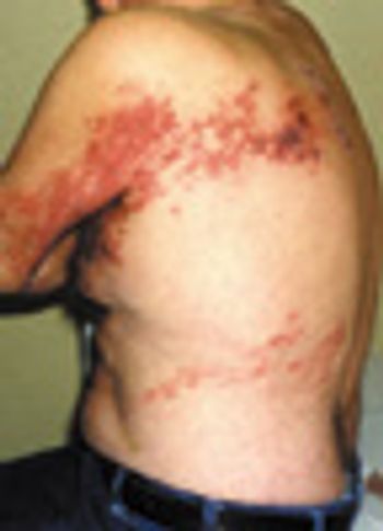
Two weeks earlier, this 66-year-old man had been hospitalized because of leftsided chest pain. However, cardiac evaluation revealed no abnormalities.
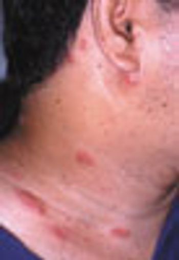
For 3 days, a 44-year-old man had several crops of tiny vesicles with raised erythematous bases on the right side of his neck and 2 elongated maculopapular lesions at the base of the neck. All of the lesions were within the C3 dermatome.
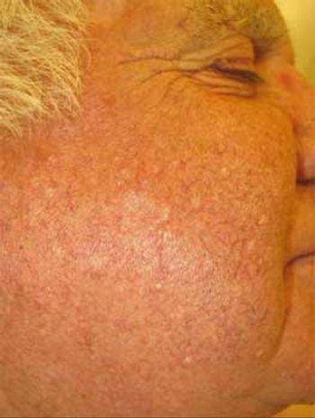
For 3 years, numerous skin-colored papules had been present on the face of a 59-year-old man. The lesions developed several months after renal transplantation and the start of immunosuppressive therapy.
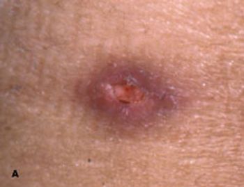
35-year-old Hispanic man presented with nonproductive cough; dyspnea; fever; and a painful, ulcerated, 1.5-cm, red-brown plaque on the left flank. He had had the lesion for 3 months and the symptoms for 1 week. The patient had grown up in Arizona, and he traveled there 4 months before the lesion arose.
Since adolescence, a 67-year old woman had had multiple nodular lesions on her body that were painful at times, particularly when pressure was applied. She reported that other family members, including her mother and brother, had similar lesions.
A54-year-old white woman presentswith extremely tender,firm lesions on the right hip and legsthat have been increasing in size andnumber over the past few months.
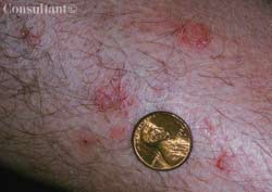
A 6-year-old girl (A) and an 11-year-old boy (B) each presentedfor evaluation of an erythematous, pruritic, papularrash that developed after swimming in a Wisconsin lake.Each child was otherwise completely healthy.
This 10-year-old boy presented forevaluation of a rash that developedduring a spring vacation on Florida’sAtlantic coast. After he had beenswimming in the ocean, a pruritic,erythematous, papular rash developedon his trunk, axillae, and groin. Approximately24 hours after the onsetof the rash, he experienced malaise,chills, and a sore throat. His past medicalhistory was unremarkable. Hehad been fully immunized and hadhad varicella infection.

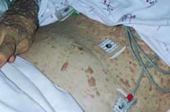
A 43-year-old woman presented to the emergency department with a 4-day history of worsening erythema, swelling, and pruritus that developed on the face and progressed to the abdomen, back, and lower legs. In the past 2 to 3 days, fluid-filled blisters had arisen, followed by skin sloughing; the patient also reported subjective fevers. Another physician had prescribed naproxen for back pain 6 days earlier. The patient had a history of asthma, with rare inhaler use, and depression, for which she had taken citalopram for 2 years.
A 72-year-old woman presented for evaluation of a large swelling onher dorsal wrist, which had been present for 2 years. The protuberancewas not painful or tender; the patient was otherwise in goodhealth.
For 3 days, a 47-year-old woman had a painful red swelling on her finger.The patient--a cellist--had tried to lance the lesion at home, but itprogressively worsened and was now “throbbing.” She denied fever andnail biting.
Swelling of the lower legs broughtthis 57-year-old woman to a familypractice clinic. She had a history ofhyperthyroidism with weight loss,tachycardia, and anxiety. This conditionwas confirmed with blood testsand radioactive iodine uptake testing.
A mildly painful, nonpruritic rash on the forearms and legs prompted a 42-year-old man to go to the emergency department. The patient noted the rashwhen he awoke that morning. He had had joint pain and fever for the past7 days and generalized malaise with chills that began about 3 days earlier.He had no significant medical history.
Hyperpigmentation is seen on the cheeks and eyelids of a 36-year-old woman.She became hyperthyroid at age 19 years, with accompanying exophthalmosand hyperpigmentation, following the birth of her first child. Thyroidectomywas carried out at that time, and the patient has been receiving thyroid replacementtherapy ever since. The hyperpigmentation, an uncommon accompanimentof hyperthyroidism, has persisted.
ABSTRACT: Acute scrotal pain, a high-riding testicle, and the absence of the cremasteric reflex on the affected side signal testicular torsion-a surgical emergency. The pain associated with torsion of the appendix testis is usually of gradual onset and is exacerbated by movement. The tenderness is often localized over the infarcted appendix, and the infarction may be visible through the scrotal skin (the "blue dot sign"). Pain associated with epididymitis is usually gradual in onset; the patient may complain of dysuria, increased frequency, and discharge, particularly if the causative pathogen is Chlamydia trachomatis or Neisseria gonorrhoeae. Hydroceles are smooth and nontender, and the scrotum transilluminates. If the scrotum does not transilluminate and compression of the fluid-filled mass toward the external ring completely reduces the mass, then a hernia is the more likely diagnosis. A patient with a varicocele typically complains of a sensation of heaviness and of "carrying a bag of worms."
A painful rash suddenly developed on the chest wall of an otherwise healthy 8-year-old girl. Examination of the rash revealed grouped vesicles with an erythematous base in a linear distribution along the T5 dermatome. The child had not been vaccinated with varicella vaccine and had had chickenpox 3 years earlier.
For more than 20 years, a 55-year-old man had a faintly erythematous, papulosquamousrash with arciform borders on his groin and waistline. The rashhad been treated with a variety of medications. Topical and oral antifungalsand antibiotics and topical corticosteroids had been used but to no avail. Nolaboratory tests had been performed.
For 1 year, a 30-year-old man had an intermittent rash that was confined to thearea of his jockey shorts; no other part of the body was affected. The patientreported that the pruritic eruption arose and disappeared spontaneously andwas more prominent during the heat of summer.
A 50-year-old man had a long-standing rash on both soles. The patient’s toenails were yellow and dystrophic. These physical findings strongly suggested moccasin-variety tinea pedis and onychomycosis.
For years, a 39-year-old man had an eruption on his hand, which seemed to becontrolled with topical corticosteroids. The patient was a rancher.
Advertisement
Advertisement
Trending on Patient Care Online
1
From Amyloid Clearance to Daytime Function: Why Sleep Quality Matters for Brain Health
2
ACOG Releases New Guidance on HIV Screening and Prevention
3
2026 Dyslipidemia Guideline Expands Statin Eligibility to 21.5 Million More US Adults
4
Compulsive Smartphone Use Linked to Depressive Symptoms in Older Adults
5