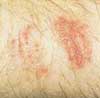
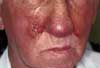
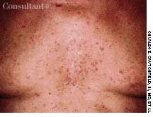
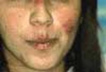
July 24th 2025
The nonsteroidal topical pan-JAK inhibitor provides a potent treatment option for adults with CHE who have not responded to corticosteroids or cannot tolerate them.
Cutaneous Signs of Vascular Disorders: Idiopathic Leukocytoclastic Vasculitis
April 2nd 2004A 16-year-old girl was bothered byankle pain and “red spots” on herlower legs. These symptoms clearedin a few days without treatment. Sixweeks later, after returning from anall-day outing at a fair, she noticedthat the spots had reappeared (A)and hemorrhagic lesions had developedon the right ankle (B) and leftheel (C). After removing her shoes,the teenager felt severe pain in bothankles, particularly the right.
Cutaneous Signs of Vascular Disorders: Atrophie Blanche
April 2nd 2004A 57-year-old man with a history of venous stasis leg ulcerationwondered about the “white spots” on his leg. Thecondition is atrophie blanche, which manifests as smooth,ivory-white macules and plaques of sclerosis stippled withtelangiectasia that often are surrounded by mild to moderatepigmentation.
Cutaneous Signs of Vascular Disorders: Carotid–Cavernous Sinus Fistula
April 2nd 2004Redness, irritation, and diplopia developedover 2 to 3 weeks in a 55-yearoldman’s left eye (A). The injectionworsened and was unresponsive toeye drops. Ptosis, mild proptosis, andelevated intraocular pressure developed.A bruit was auscultated overthe affected eye.
Cutaneous Signs of Vascular Disorders: Polycythemia Vera
April 2nd 2004Four months after a patchy, macular,erythematous spot erupted on thedorsum of a 63-year-old woman’s leftfoot, the area became ulcerated,tender, and painful. The 1.2-cm ulcerwas covered by a hemorrhagic crustsurrounded by a cyanotic reticulardiscoloration of the skin.
Various Manifestations of Rheumatic Disorders: Case 6 Progressive Systemic Sclerosis
March 2nd 2004For several months, a 70-year-old woman had had dysphagia,mild dyspnea on exertion, and the Raynaud phenomenon.Her skin was waxy and edematous; 2- to 10-mm pinkishspots had appeared on her fingers, palms, and oral mucousmembrane over the past 2 weeks. These disappearedcompletely with pressure. Subcutaneous calcific depositswere present on the extensor surfaces of the forearms.
Various Manifestations of Rheumatic Disorders: Case 5 Rheumatoid Nodules
March 2nd 2004A 65-year-old woman, who was confined to a wheelchairbecause of severe rheumatoid arthritis, was concernedabout nodules that had erupted on her fingers and handsduring the previous 3 weeks (A). Her medical historyincluded colon cancer, chronic renal insufficiency, anemia,and hypertension. The nonpruritic nodules were painfulwhen they began to form under the skin; however, oncethey erupted, the pain disappeared.
Various Manifestations of Rheumatic Disorders: Case 3 Rheumatoid Vasculitis
March 2nd 2004Ten weeks before presentation, this55-year-old woman noticed decreasedsensation in her feet and a bluish discolorationof her toes. These symptomsprogressed rapidly, and pain andcoldness in both feet increased in intensity.Her feet subsequently becamegangrenous. Her seropositive arthritishad been diagnosed about 6 yearsearlier. The disease had been wellcontrolled until about 10 weeks beforethis photograph was taken.
Various Manifestations of Rheumatic Disorders: Case 2 Heberden Nodes
March 2nd 2004Bilateral swelling and pain in the distal interphalangeal (DIP) joints for severalmonths brought this 65-year-old woman to her physician. She complained alsoof stiffness in the region of the DIP joints when she arose in the morning andafter short periods (less than 15 minutes) of inactivity. A history such as this,in conjunction with the appearance of the patient’s hand, is typical of Heberdennodes, which are a manifestation of osteoarthritis (OA).
Tinea Types: Common Dermatophyte Infections Case 3 Bullous Tinea Pedis
February 1st 2004A 24-year-old man presented for evaluation of pruritic vesicles on both feet.Ten days earlier, dyshidrotic eczema had been diagnosed by another physicianwho prescribed triamcinolone ointment. The patient reported that the footeruption worsened after the topical medication was applied.
Tinea Types: Common Dermatophyte Infections Case 2 Moccasin-Variety Tinea Pedis
February 1st 2004A 70-year-old man first noticed thisskin condition when he returned fromthe South Pacific at the end of WorldWar II. Over the years, the rash hasitched only occasionally; however,during a recent spate of hot weather,the eruption became highly pruritic.Applications of an over-the-counter1% hydrocortisone ointment exacerbatedthe condition
Tinea Types: Common Dermatophyte Infections Case 9 Tinea Faciei
February 1st 2004An eruption on the face of a 49-year-old woman had been misdiagnosed as astaphylococcal infection; the rash failed to respond to oral and topical antibiotics.A mid-potency topical corticosteroid also had been tried, but the eruptionworsened.
Tinea Types: Common Dermatophyte Infections Case 8 Tinea Capitis
February 1st 2004The parents of a 3-year-old girl sought evaluation of their daughter’s hair loss.During the past several months, a large patch of alopecia with scaling had developed.The differential diagnosis included seborrhea, trichotillomania, andtinea capitis.
Tinea Types: Common Dermatophyte Infections Case 6 Negative-Culture Tinea Corporis
February 1st 2004A 49-year-old man was concerned about a right flexor forearmlesion that had been increasing in size for 6 weeks.The light pink, well-demarcated, 5-cm, circular lesion featuredslight peripheral elevation with ulceration, crusting,and a relatively clear central area. A culture of materialfrom the lesion was negative for fungi. A potassium hydroxideevaluation was not performed.
Tinea Types: Common Dermatophyte Infections Case 4 Tinea Manuum and Tinea Unguium
February 1st 2004For about 4 months, a very dry, diffuse,fine scaly, asymptomatic eruptioncovered the palms of a 28-yearoldman; several fingernails weredystrophic bilaterally as well. Beforethe onset of this condition, bilateralonychomycosis of the toenails hadbeen diagnosed. The toenails had notbeen treated and were still affectedat the time of presentation. Branchinghyphae were seen on a potassiumhydroxide preparation of a fingernailcutting. The patient had tinea manuumand tinea unguium
Is There a Medical Explanation?
February 1st 2004A 13-year-old girl of African American descent is brought to the pediatrician’s office becauseof a lesion on her neck. The girl’s mother had telephoned the office before the visit, statingthat the lesion resembled a blister at first, but now looked like a burn.
Images of Malignancy: Case 4 Peritoneal Carcinomatosis
January 2nd 2004Vague abdominal pain, malaise, anorexia,and the loss of 10 lb in 2months prompted a 65-year-old manto seek medical evaluation. A yearearlier he had undergone surgery forstage III carcinoma of the sigmoidcolon. Because metastases to thelymph nodes were found in the resectedcolon, the patient was given postoperativechemotherapy. Histologicexamination revealed poorly differentiatedadenocarcinoma.
Images of Malignancy: Case 3 Metastases to the Scalp
January 2nd 2004An 85-year-old man was admitted to the hospital with acough and shortness of breath of 1 week’s duration anda fever and increased sputum production for 2 days. Hishistory included renal cell carcinoma and metastatic renalcancer for 2 years. The patient had smoked cigarettesfor 30 years. He had lost 30 lb during the last few months.A chest film revealed pneumonia of the right lowerlobe. Metastatic nodules were noted on the scalp; extensivelung, bone, and brain metastases also were found.
Images of Malignancy: Case 2 Papillary Carcinoma of the Thyroid
January 2nd 2004A 40-year-old man was concerned about an enlarging painlessmass on the right side of his neck that had been presentfor 6 months (A). The patient reported no other healthproblems; his medical history was unremarkable, and hewas taking no medications.There was no family or personal history of thyroiddisease or of exposure to radiation. Thyroid function testresults were within normal limits. A chest film revealed nopathology.
Peripheral Arterial Disease: Tips on Diagnosis and Management
January 1st 2004Signs and symptoms that strongly suggest peripheral arterial occlusive disease include diminished or absent pedal pulses, a unilaterally cool limb, and atrophic skin that is shiny and hairless. An ankle-brachial index of less than 0.5 suggests multisegment disease. Management goals are to decrease functional impairment, treat underlying atherosclerosis, and control risk factors. Smoking cessation is imperative. A graduated walking program is a mainstay of treatment and is associated with greater improvement in pain-free walking than is drug therapy. Surgery and percutaneous intervention are generally reserved for patients with lifestyle-limiting claudication, ischemic pain at rest, tissue loss, or gangrene.
Blistering Skin Condition: Hailey-Hailey Disease
December 1st 2003Several times a year a rash erupts on the chest, axillae, and neck of a 41-year-old woman. Her father and siblings have a similar history. A biopsy of the affected skin confirmed the suspected diagnosis of benign familial pemphigus, which is also called Hailey-Hailey disease.