

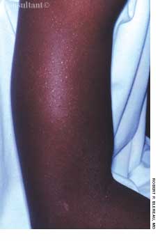


The continuous use of a corticosteroid cream briefly relieved the pruritus of anannular, papulosquamous eruption on the left anterior thigh of a 50-year-oldwoman. The lesion was present for 6 months and grew larger with applicationof the topical corticosteroid.
Dermatology
Latest News
Advertisement
Advertisement
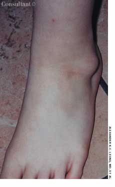
For several months, a 49-year-old woman has had asymptomatic loss of pigmenton her shins. She has no history of injury to the area.What is your clinical impression?
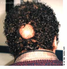
During a routine physical examination, a white forelock was noted on a 54-year-old man. The patient stated that the discolored patch of hair had been present since adolescence. Other than mild hearing loss, he had no significant personal or family medical history.
A 46-year-old man with diabetes presented for evaluation of gradual fingernail deterioration, which had failed to respond to several courses of griseofulvin and a recent 3-month course of daily terbinafine. The patient-who worked as a bartender-was otherwise healthy.

For several weeks, a 78-year-old woman had an intensely pruritic, diffuse, raised, slightly scaly, erythematous rash that persisted despite the use of several over-the-counter topical medications (such as hydrocortisone and clotrimazole cream). Since her last visit about 3 months earlier for a blood pressure reading, she had been well except for 2 episodes of night sweats.
The American Cancer Society predicts that 55,100 cases of melanoma will be diagnosed in 2004.1 More than 1 million new cases of basal cell and squamous cell carcinoma are expected.2
Sorting through the myriad of causes of soft tissue infections can be a daunting diagnostic challenge. While much is written about empiric treatment of skin and soft tissue infections, it is important to make a correct diagnosis, since clinical findings in common versus exotic and mild versus life-threatening infections have significant overlap. Historical information, such as the temporal progression of signs and symptoms, travel history, animal exposure, age, occupation, bite history, underlying diseases, and lifestyle, is important in focusing the differential diagnosis toward specific causes. Still, clinical assessment is frequently not sufficient and laboratory tests, radiographic imaging, and surgical intervention may be necessary to establish a specific diagnosis and to provide the rationale for definitive management.
A 52-year-old man complains of nausea, fever, and malaise following a 2-day diarrhealillness that developed at the end of a family vacation in New England.Two family members suffered a similar illness, characterized by watery diarrhea.Symptoms developed in all who were affected within 24 hours of eatinghamburgers at a local restaurant.
A 70-year-old man who had just completeda course of trimethoprim-sulfamethoxazolefor a urinary tract infectionpresented with palpable purpuraand cutaneous erosions of acute onseton his legs (A). He also had massivescrotal edema and purpura (B).
Painful blue toes developed in a 72-year-old woman with coronary artery andperipheral vascular disease after she underwent angiography.
A 16-year-old girl was bothered byankle pain and “red spots” on herlower legs. These symptoms clearedin a few days without treatment. Sixweeks later, after returning from anall-day outing at a fair, she noticedthat the spots had reappeared (A)and hemorrhagic lesions had developedon the right ankle (B) and leftheel (C). After removing her shoes,the teenager felt severe pain in bothankles, particularly the right.
A 57-year-old man with a history of venous stasis leg ulcerationwondered about the “white spots” on his leg. Thecondition is atrophie blanche, which manifests as smooth,ivory-white macules and plaques of sclerosis stippled withtelangiectasia that often are surrounded by mild to moderatepigmentation.
Redness, irritation, and diplopia developedover 2 to 3 weeks in a 55-yearoldman’s left eye (A). The injectionworsened and was unresponsive toeye drops. Ptosis, mild proptosis, andelevated intraocular pressure developed.A bruit was auscultated overthe affected eye.
Sudden pain and a pulsatile swellingof the right upper medial thigh concernedan 80-year-old woman (A).Coronary angioplasty had been performedthrough this site 5 weeksearlier.
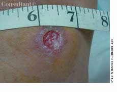
Four months after a patchy, macular,erythematous spot erupted on thedorsum of a 63-year-old woman’s leftfoot, the area became ulcerated,tender, and painful. The 1.2-cm ulcerwas covered by a hemorrhagic crustsurrounded by a cyanotic reticulardiscoloration of the skin.
ABSTRACT: Because physical findings are an unreliable indicator of deep venous thrombosis (DVT), the diagnosis is based on the presence of clinical risk factors and the results of noninvasive tests, such as duplex ultrasonography and impedance plethysmography. Contrast venography is considered the gold standard for the diagnosis of DVT. Uncomplicated DVT is managed with low molecular weight heparin followed by warfarin. When DVT is complicated (eg, by pregnancy or by evidence of pulmonary embolism), the patient is treated with intravenous heparin; the dosage is adjusted to achieve an activated partial thromboplastin time 3 times control. Chronic venous insufficiency is the most common cause of leg ulcers. Treatment goals include reduction of edema, relief of pain, ulcer healing, and prevention of recurrence. Leg elevation and multilayer elastic compression dressings are the mainstays of therapy. Compression dressings are continued until ulcers heal; graded compression stockings are worn to prevent recurrence. Pentoxifylline, 400 mg 3 times a day, is an effective adjunct to compression bandaging. Large or slow-healing ulcers may require skin grafts.
During the past week, a rash on theright thumb and forefinger of a 4-yearoldboy has progressed to involve theentire arm.
For several months, a 70-year-old woman had had dysphagia,mild dyspnea on exertion, and the Raynaud phenomenon.Her skin was waxy and edematous; 2- to 10-mm pinkishspots had appeared on her fingers, palms, and oral mucousmembrane over the past 2 weeks. These disappearedcompletely with pressure. Subcutaneous calcific depositswere present on the extensor surfaces of the forearms.
A 65-year-old woman, who was confined to a wheelchairbecause of severe rheumatoid arthritis, was concernedabout nodules that had erupted on her fingers and handsduring the previous 3 weeks (A). Her medical historyincluded colon cancer, chronic renal insufficiency, anemia,and hypertension. The nonpruritic nodules were painfulwhen they began to form under the skin; however, oncethey erupted, the pain disappeared.
A 76-year-old woman complained of progressive proximalmuscle weakness; achy pain in the buttocks, thighs, andcalves; and lilac discoloration of her eyelids, cheek, nose,knuckles, and fingernails.
Ten weeks before presentation, this55-year-old woman noticed decreasedsensation in her feet and a bluish discolorationof her toes. These symptomsprogressed rapidly, and pain andcoldness in both feet increased in intensity.Her feet subsequently becamegangrenous. Her seropositive arthritishad been diagnosed about 6 yearsearlier. The disease had been wellcontrolled until about 10 weeks beforethis photograph was taken.
Bilateral swelling and pain in the distal interphalangeal (DIP) joints for severalmonths brought this 65-year-old woman to her physician. She complained alsoof stiffness in the region of the DIP joints when she arose in the morning andafter short periods (less than 15 minutes) of inactivity. A history such as this,in conjunction with the appearance of the patient’s hand, is typical of Heberdennodes, which are a manifestation of osteoarthritis (OA).
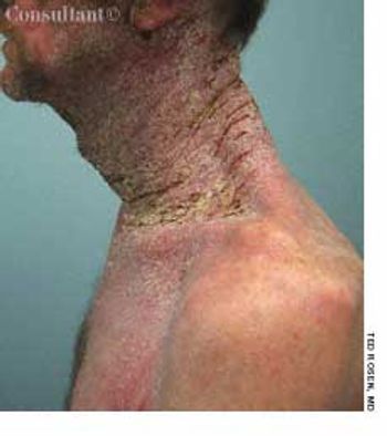
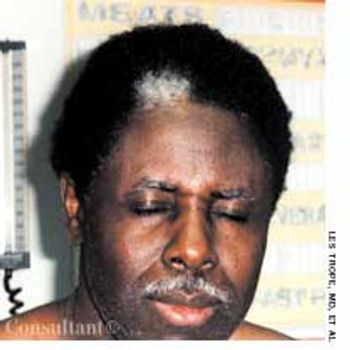
A52-year-old white man presented with a pruritic eruption on the neck of 3 months’ duration. The rash had not responded to a potent topical corticosteroid prescribed by another practitioner for the presumed diagnosis of eczema. The patient reported no current health problems. His history included a pubic louse infestation and several episodes of uncomplicated urethral gonorrhea. He readily admitted to having unprotected sexual intercourse with prostitutes.
A 24-year-old man presented for evaluation of pruritic vesicles on both feet.Ten days earlier, dyshidrotic eczema had been diagnosed by another physicianwho prescribed triamcinolone ointment. The patient reported that the footeruption worsened after the topical medication was applied.
A 70-year-old man first noticed thisskin condition when he returned fromthe South Pacific at the end of WorldWar II. Over the years, the rash hasitched only occasionally; however,during a recent spate of hot weather,the eruption became highly pruritic.Applications of an over-the-counter1% hydrocortisone ointment exacerbatedthe condition
Advertisement
Advertisement
Trending on Patient Care Online
1
ACOG Releases New Guidance on HIV Screening and Prevention
2
From Amyloid Clearance to Daytime Function: Why Sleep Quality Matters for Brain Health
3
2026 Dyslipidemia Guideline Expands Statin Eligibility to 21.5 Million More US Adults
4
FDA Accepts Cefiderocol sNDA for Pediatric Gram-Negative Infections
5