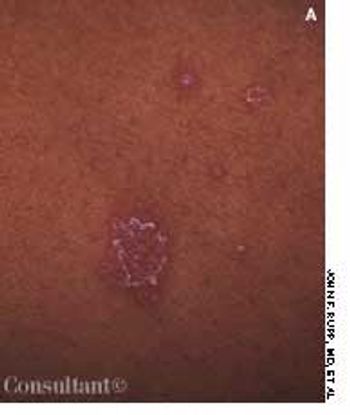
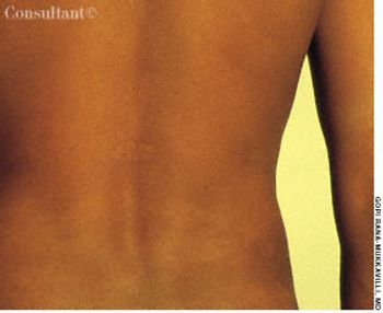
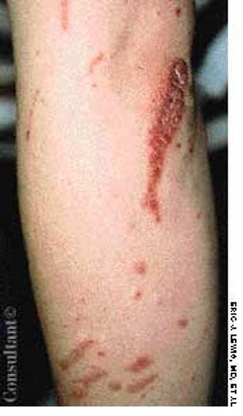
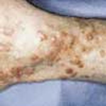
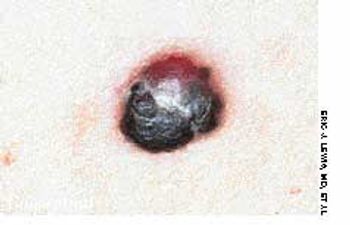
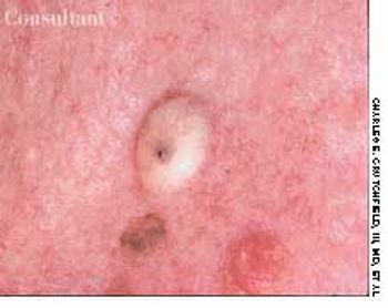
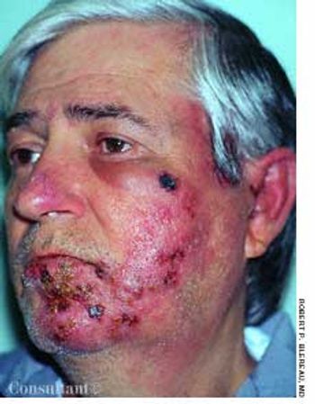
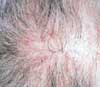
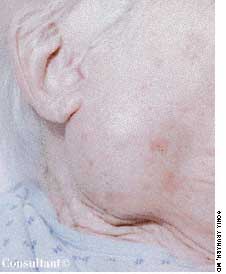
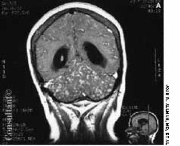
Dermatology

Latest News


CME Content


For 3 weeks, a 14-year-old boy had been aware of an enlarging lesion on the back of his hand. He recalled no trauma to the affected area. Further questioning by Dr D. Keith Cobb of Savannah, Ga, revealed that a 4-mm verruca, or wart, had been removed from the same site 6 months earlier with cryosurgery by a different physician.

Redness and swelling of the left cheek, chin, and ear bothered a 51-year-old man. The initial diagnosis was cellulitis and/or allergic dermatitis; oral amoxicillin/clavulanate, 500 mg tid, and a low- to medium-potency corticosteroid cream, alclometasone, were prescribed. Within 1 to 2 days, pimples emerged in the reddened areas and rapidly crusted.

Hyperpigmentation involving the neck developed in this 8-year-old obese girl. The affected area resembled unwashed skin. The patient had worn a uniform to school-a jumper and blouse with a collar-for 6 months.

A 6-month-old girl presented with a reddish mass on the abdomen that was not apparent at birth and was first noted when the child was 1 month old. The lesion-which was asymptomatic-started to shrink and fade when the child was 3 years old. A year later, the color was very faint. When the child was 6 years old, the lesion was hardly visible.

For several weeks, a 78-year-old woman had an intensely pruritic, diffuse, raised, slightly scaly, erythematous rash that persisted despite the use of several over-the-counter topical medications (such as hydrocortisone and clotrimazole cream). Since her last visit about 3 months earlier for a blood pressure reading, she had been well except for 2 episodes of night sweats. For several years, she had been taking levothyroxine and reserpine/hydrochlorothiazide; about 6 months ago, valsartan/hydrochlorothiazide had been prescribed.

A mildly painful, nonpruritic rash on the forearms and legs prompted a 42-year-old man to go to the emergency department (ED). The patient noted the rash when he awoke that morning. He had had joint pain and fever for the past 7 days and generalized malaise with chills that began about 3 days earlier. He had no significant medical history.

A male infant was delivered at term to a 24-year-old woman. The pregnancy had been uncomplicated; the vaginal delivery was normal. Apgar scores were 7 at 1 minute and 9 at 5 minutes. Birth weight was 3020 g (6 lb 11 oz); length, 51 cm (20 in); and head circumference, 36 cm (14 in).

This lesion consists of a dilated central feeding arteriole and smaller radiating branches that together suggest a spider's body and legs. The lesion blanches when pressure is applied. Pulsations visible in larger nevi are evidence of the arterial source of the lesion.

A 40-year-old man was hospitalized with the superficial, reddening, and painful facial lesion seen here. Its borders were sharp, and it had developed rapidly. The patient had a temperature of 40°C (104°F) and chills. His erythrocyte sedimentation rate was elevated, and he had neutrophilic leukocytosis.

After 1 week of scratching a pruritic, reddish rash on a swollen hand and enduring a “burning sensation,” a 43-year-old man visited his physician. The patient worked as a meatpacker. He had no other medical conditions, took no medications, and had no known drug allergies.

A 68-year-old man presented with a sudden-onset, 2.5 × 2-cm, rock-hard, erythematous, nontender nodule on the right side of the chest. A dense mat of telangiectases surrounded the solitary lesion. The remainder of the cutaneous examination was unremarkable.

Tortuous, dilated varicosities; multiple smaller caliber abnormal perforating vessels; and chronic brawny edema of chronic venous insufficiency (CVI) were seen on a 70-year-old man's left leg. He reported that the edema and discoloration had worsened over the last 15 years. The brawny edema stopped just above the ankle, indicating that compression by the patient's sock controlled the signs and symptoms of CVI.

A 60-year-old woman was referred by her gynecologist because of a lesion on the buttocks of which the patient first became aware when she noticed blood on her underwear. Physical examination revealed an irregularly pigmented and slightly eroded asymmetric plaque. Examination with a magnifier highlighted a slightly rolled border, from which a shave biopsy was performed.

Asteatosis

Erythematous, scaly lesions with double-edged borders had been present on a 14-year-old boy's left upper arm and lower legs for about a year. The lesions were occasionally pruritic, and some resembled ringworm. At times, fine yellow crusting suggestive of impetigo was present. The boy took very hot baths and showers.

Tinea Faciei

An eruption on the face of a 49-year-old woman had been misdiagnosed as a staphylococcal infection; the rash failed to respond to oral and topical antibiotics. A mid-potency topical corticosteroid also had been tried, but the eruption worsened.

Mongolian spots are benign, congenital blue-gray macules or patches commonly found in Asian, black, and Hispanic infants (and in 5% of white infants). They are most often located in the sacrogluteal area and the upper back. The lesions may be solitary or multiple, vary in color intensity, and frequently enlarge until about age 2, when they gradually fade and disappear. Approximately 5% persist into adulthood.

The widespread eruption of asymptomatic macules and flat, palpable, flesh-colored lesions prompted a 23-year-old woman to consult her physician. The lesions-some of which had dark centers-were concentrated on the patient's face, neck, and upper back; the palms, soles, anal mucosa, and genital areas were clear. The patient denied systemic symptoms. She was seronegative for HIV.