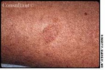























































































































































Concern about this flat, tan, atrophic, well-marginated, dime-sized lesion prompted a 44-year-old woman to seek medical advice. The lesion, which was mildly pruritic, had appeared 2 months earlier.


Concern about this flat, tan, atrophic, well-marginated, dime-sized lesion prompted a 44-year-old woman to seek medical advice. The lesion, which was mildly pruritic, had appeared 2 months earlier.

Three months ago, a 50-year-old man who was otherwise in good health noticed a hard, round nodule on his left arm. Within 2 months, similar nodules appeared all over his trunk, head, arms, and legs. The reddish purple lesions, less than 2 cm in diameter, were painless and slightly pruritic.

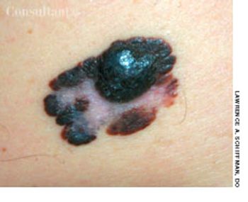
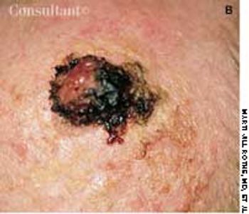
This truncal lesion was revealed when a 47-year-old man disrobed for examination after he had fallen on a sheet of ice and broken a rib. The patient-a truck driver and 30 pack-year cigarette smoker-had been aware of the enlarging lesion for about a year but had not sought medical attention because it was asymptomatic.

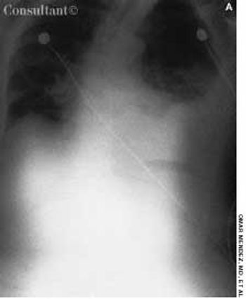
An 85-year-old white woman was brought to the emergency department (ED) with acute, severe left posterolateral chest wall pain of several hours' duration. The nonradiating pain was accompanied by shortness of breath. She denied palpitations, diaphoresis, syncope, or dizziness.

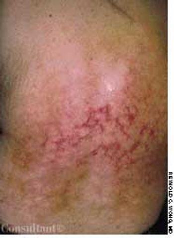
An area of mottled skin developed on the back of a 55-year-old woman who has had arthritis for several years. She often applied a heating pad to her middle and lower back for relief from arthritic pain.

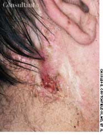
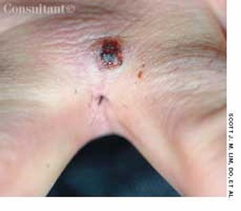
An ulcerating nodule on his left pre-auricular region worried a 64-year-old man.

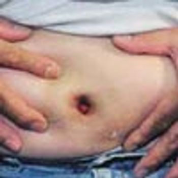
A 37-year-old man presented with a reddish nodule in the umbilical area that had been present since early infancy. The lesion had been cauterized with silver nitrate several times in the past but had not resolved. No drainage or secondary infection was noted, and the patient was otherwise in good health.

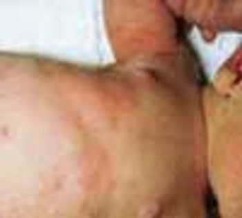
At 3 days of age, this infant was noted to have an erythematous, maculopapular rash on the face, chest, abdomen, and back that included some pustule-like lesions.

In the web space of his left hand, a 50-year-old barber had a painful cystlike lesion. The lesion had recurred intermittently, despite oral antibiotic treatment and warm compresses. The patient's father, also a barber, had a similar, more severe condition, which eventually required surgical intervention.

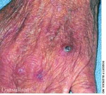
For 1 month, a 66-year-old man had had an asymptomatic lesion on the dorsum of his left hand. The flesh-colored, dome-shaped, maroon-crusted lesion measured 0.7 cm in diameter and was located over the fourth knuckle. The patient had chronic obstructive pulmonary disease but was otherwise in good health. He was seronegative for HIV.

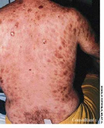
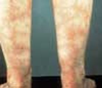
A 55-year-old woman with no significant medical history reported that diffuse erythematous, patchy, purpuric skin lesions over most of her body had been present for the past year. Applications of an over-the-counter corticosteroid cream helped control the pruritus but did not clear the skin lesions.

During a routine physical examination, a white forelock was noted on a 54-year-old man. The patient stated that the discolored patch of hair had been present since adolescence. Other than mild hearing loss, he had no significant personal or family medical history.

A previously healthy 52-year-old man had been using an antidandruff shampoo and a topical corticosteroid for a chronic scalp eruption that had been diagnosed as seborrheic dermatitis. Because the condition had not responded to this regimen, a scalp biopsy was performed; the findings were consistent with seborrheic dermatitis.

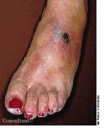
A patchy, macular, erythematous area developed on the dorsum of a 63-year-old woman's left foot 4 months ago. Because the lesion was asymptomatic and she was otherwise healthy, no workup was instituted at that time. The affected area subsequently became ulcerated, tender, and painful. The patient now had a 1.2-cm ulcer covered by a dried, hemorrhagic crust and surrounded by a livedo reticularis–like pattern. The entire area was extremely tender.

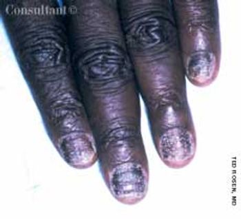
A 43-year-old man sought treatment of a “fungal nail infection” that had been present for several years. The condition had not responded to standard dosages of itraconazole, terbinafine, and fluconazole prescribed by another physician.

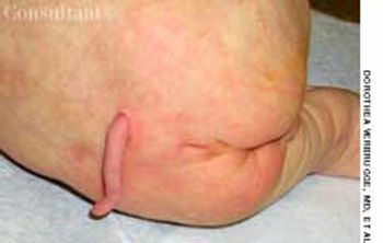
A 6-cm midline tail-like soft tissue appendage was noted on the back of this newborn infant at the level of L5. An epithelialized mid-sacrum dimple was also present inferior to the appendage. The infant otherwise appeared to be normally developed.

A pruritic lesion on the arm of a 26-year-old woman had grown to 22 cm while she was "treating" the eruption with a topical corticosteroid.

An 82-year-old man with Alzheimer's disease and atrial fibrillation was referred for evaluation of a lesion present for an undetermined period. It was initially noted 1 week earlier, at his first office visit with a geriatrician.

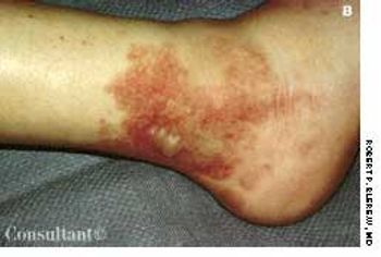
A 16-year-old girl was bothered by ankle pain and “red spots” on her lower legs. These symptoms cleared in a few days without treatment. Six weeks later, after returning from an all-day outing at a fair, she noticed that the spots reappeared and hemorrhagic lesions developed on the right ankle and left heel. After removing her shoes, the teen-ager felt severe pain in both ankles, particularly the right.

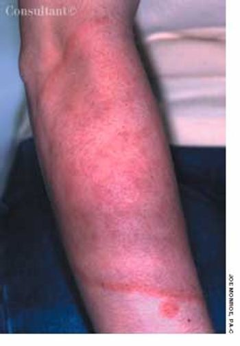
Livedo reticularis is the cutaneous manifestation most frequently associated with the antiphospholipid syndrome (APS). It manifests as a dusky, violaceous vascular discoloration with a reticulated pattern on the upper and lower extremities.

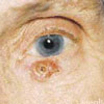
An otherwise healthy 76-year-old woman with no chronic systemic conditions requested removal of a left lower eyelid growth she feared was cancerous. The patient was correct; excision of the lesion and histologic examination confirmed basal cell carcinoma.

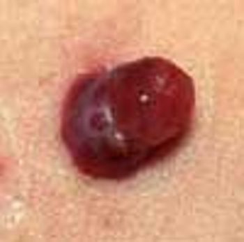
A 1 × 0.5-cm hemorrhagic, polypoid lesion that had been present for 2 months on a 13-year-old boy's left anterior chest was excised. Pathologic examination confirmed the diagnosis of pyogenic granuloma.

A 32-year-old woman became concerned about the numerous bumps on her upper face, which had been increasing in number for many years. Examination revealed clusters of 2- to 3-mm lesions-mostly monomorphic, flesh-colored papules lateral to the eyebrows and on the central forehead, inferior-medial eyelids, and upper cheeks. Some of the papules had a slight pink or yellow-brown appearance, and poorly demarcated brown patches (melasma) were noted on the cheeks.

Lichen planus is characterized by flat-topped, polygonal, purple pruritic papules that have a predilection for flexor aspects of the wrists and forearms, sides of the neck, thighs, shins, and lower back. Lesions on the oral mucosa appear as white, lacy patches.

This is a common infectious problem, especially among school-age children. In many cases, there are well-defined areas of hair loss that can mimic alopecia areata.