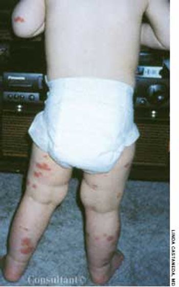





















































































































































For 1 week, a 2-year-old boy has hadasymmetric erythematous patcheson the anterior trunk. There is nofamily or personal history of eczema.His mother has not used new soapsor detergents recently; there are nopets in the household.

For 1 week, a 2-year-old boy has hadasymmetric erythematous patcheson the anterior trunk. There is nofamily or personal history of eczema.His mother has not used new soapsor detergents recently; there are nopets in the household.

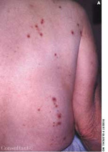
Highly pruritic, 2- to 4-mm, papular lesions with central ulceration erupted on the back of a 66-year-old woman. She had had 2 similar outbreaks in the past. The patient was taking conjugated estrogens, alprazolam, and alendronate.

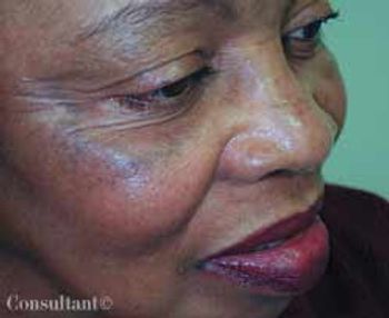
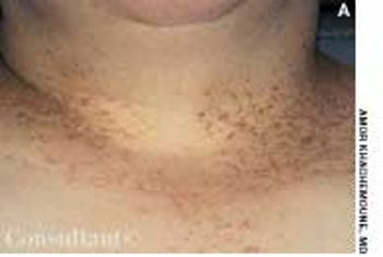
A 58-year-old black woman sought evaluation of areas of increased pigmentation on her cheeks that had been present for many years. Bleaching agents did not lighten the area; new, non–nickel-containing eyeglass frames had no effect. A 3-week course of halobetasol cream failed to fade the hyperpigmentation.

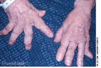
A 76-year-old woman had a 40-year history of rheumatoid arthritis (RA). She had repeatedly refused treatment with disease-modifying drugs, including methotrexate. Nodules began to develop 15 years after the initial diagnosis; they recurred after surgical removal.

Cutaneous manifestations develop inapproximately 30% of persons withdiabetes. Premature atherosclerosisis a common complication of thedisease and can cause peripheral infarction,ulceration, and necrosis.

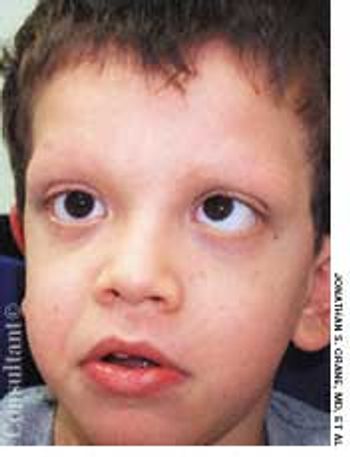
Syndrome The mother of a 7-year-old boy with cardiofaciocutaneous syndrome sought treatment for the cutaneous aspects of her son’s disease. Dry skin and keratosis pilaris of the upper outer arms were noted. Cardiofaciocutaneous syndrome- a rare autosomal dominant genetic disorder-had been diagnosed when the child was 3 years old.

A 42-year-old woman presented with a papular eruption that first appeared during childhood. Severe pruritus, which worsened during the summer, accompanied the lesions. The patient reported that family members, including her mother and brother, had a similar skin problem. A biopsy of a characteristic lesion on the patient’s chest revealed diffuse acantholytic dyskeratosis, which confirmed the clinical diagnosis of Darier’s disease.

This patient with long-standing insulin-dependent diabetes has difficultyclosing his hand because of “tight skin.” Diabetes is the cause: this findingoccurs more often in persons with microvascular complications, such asretinopathy and nephropathy. The condition may occur even in patients withwell-controlled diabetes.

This huge blister developed spontaneously on the foot of a woman withdiabetes mellitus. There was no area of redness around the blister, whichnormally occurs with burns and inflamed lesions. Spontaneous blisters andother skin manifestations are common in persons with diabetes.

Numerous brown spots on his leg brought this man with long-standing diabetes mellitus to his physician. He thought the spots were related to trauma.

This patient with long-standing insulin-dependent diabetes mellitus has difficulty in closing his hand because the skin is "very tight."

Numerous brown spots on the leg of a man with longstandingdiabetes are diabetic dermopathy. This is amicroangiopathy of the small arterioles of the legs, whichresults in atrophic brown papules.

The atrophic patches on the lower legs of this 47-year-old woman who has haddiabetes mellitus for over 20 years are those of necrobiosis lipoidica diabeticorum.These areas involve degeneration of collagen and elastic fibers in thelower dermis and changes in blood vessel walls. The lesions usually begin assingle or multiple elevated reddish nodules, most commonly in the pretibialarea. Over time, they expand and coalesce into distinctive brownish yellowpatches. These areas may be somewhat tender, but as they spread, theyfrequently become painless-unless they ulcerate. Because the involved skinis fragile, ulcers can form after any minor trauma.

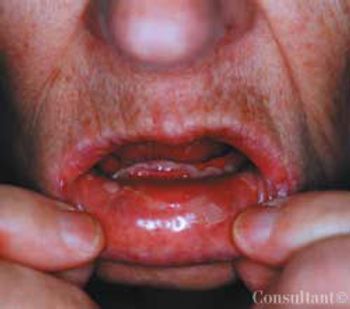
Levofloxacin, 500 mg/d, had been prescribed for a 74-year-old woman who had a urinary tract infection. The patient had type 2 diabetes and hypertension. She was allergic to sulfa drugs. Two hours after taking the first oral dose of the antibiotic, painful blisters developed on the lower lip and soft palate.

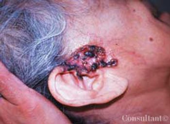
A 75-year-old Hispanic woman presented with a slowly growing, asymptomatic facial lesion of about 3 years’ duration.

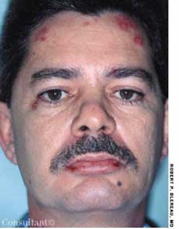
Numerous plaques, some with yellow crusting and central scarring, had erupted primarily on the face and neck of a 46-year-old man. A single lesion had developed on his left elbow as well. The lesions were initially diagnosed as impetigo, but they failed to resolve after 2 courses of oral cephalexin.

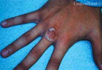
For 3 weeks, a 14-year-old boy had been aware of an enlarging lesion on the back of his hand. He recalled no trauma to the affected area. Further questioning by Dr D. Keith Cobb of Savannah, Ga, revealed that a 4-mm verruca, or wart, had been removed from the same site 6 months earlier with cryosurgery by a different physician.

Concerned about a lesion between her eyes, a 91-year-old woman sought medical evaluation. She had not seen a physician for 23 years. The patient was strong-willed and alert, with no evidence of Alzheimer disease. A huge goiter was evident.

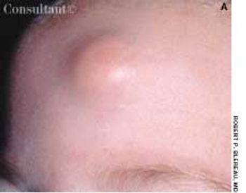
A 0.5-cm mass with overlying erythema on the forehead of a 5-monthold girl was first noticed after she had been hit on the head with a rattle. Excision of the mass was deferred because it was considered to be a hemangioma.

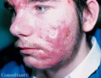
For the past 3 years, comedones, papules, pustules, and nodules had been erupting on the face of a 16-year-old boy. Acne vulgaris had been diagnosed. Topical tetracycline cream and oral tetracycline were used without success.

Painful erosions developed on the sole of a 14-year-old girl's foot several weeksearlier. Within the last few days, the condition has spread to the other sole.The patient is otherwise healthy and takes no medications. She enjoys playingsoccer and has no history of trauma.

Four pink nodules appeared in a linear array on the proximal extensor right forearm of a 77-year-old man. The asymptomatic lesions, which ranged from 0.5 to 1.0 cm in diameter, had been present for 2 months.

Is soaking the feet in an aluminum salt solution an effective therapy fordyshidrosis?

A 49-year-old woman was admitted tothe hospital with a high fever ofabrupt onset, rigor, headache, myalgias,and profound prostration. Hertemperature was 41ºC (105.8ºF);blood pressure, 60/40 mm Hg; respirationrate, 30 breaths per minute;and pulse rate, 130 beats per minute.

A 55-year-old woman had a 10-day historyof a nonproductive cough and apainful rash on both palms for 5 days.The eruption subsequently spreadover her entire body.