
|Articles|July 31, 2013
GI Lesions and Gut Feelings-A Photo Essay
Here: 5 Tips about Crohn disease, Pseudopolyps, GI Cancer, and other GI Maladies
Advertisement
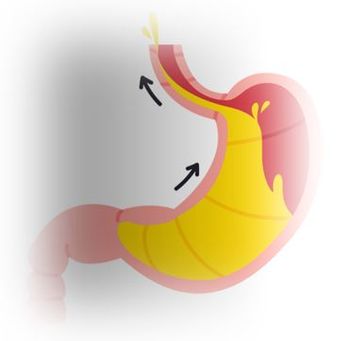
Patients with inflammatory bowel disease-especially those with
Click here for the next image
A 17-year-old man with an 18-month history of “a nervous stomach” awoke with severe pubic area pain. His temperature was 97.1°F; physical exam revealed localized tenderness at McBurney point. The WBC count was 20,300/μL, with 91% neutrophils and 9% lymphocytes. Abdominal ultrasonography demonstrated a bull’s eye typical of
Click here for the next image
A 26-year-old man presented to the emergency department with progressively worsening generalized fatigue and weakness that had been present for the past few months. A family history was notable for colon cancer in his father and grandfather. Results of laboratory studies were significant for iron deficiency anemia. Colonoscopy revealed innumerable polyps carpeting the mucosa from the rectum to the cecum. The diagnosis:
Click here for the next image
For 6 weeks, a 29-year-old previously healthy man had between 10 and 15 episodes daily of small-volume bloody diarrhea with intermittent paraumbilical pain. Colonoscopy revealed diffuse ulceration with loss of vascularity and mucosal surfaces that extended from the rectum to the cecum. Pseudopolyps-distinct, irregular, raised areas of normal-appearing mucosa-were noted among the areas of friability, fibrous stranding, and ulceration. Pseudopolyps, which represent a combination of reactive hyperplasia and mucosal ulceration, are not uncommon in severe or chronic
Click here for the next image
Click here to return to the first image.
Newsletter
Enhance your clinical practice with the Patient Care newsletter, offering the latest evidence-based guidelines, diagnostic insights, and treatment strategies for primary care physicians.
Advertisement
Related Content
Advertisement


Latest CME


Advertisement
Advertisement
Trending on Patient Care Online
1
First Oral Film Treatment for Erectile Dysfunction in Men Gains FDA Approval
2
Long-Term Data Support Sustained Bimekizumab Response in Hidradenitis Suppurativa
3
Weekly Dose Podcast: New Obesity Data, Insulin Guidance, and Mental Health Screening
4
Topline Phase 2 Data Show Roflumilast Cream Improves Atopic Dermatitis in Infants as Young as 3 Months
5