Dermatitis on the Hands and Chest
A 12-year-old boy with a history of atopy complained of pruritus and severe dryness of the hands. Over-the-counter moisturizers failed to resolve the condition. The patient did not wash his hands frequently and had no hobbies that exposed him to environmental irritants or allergens.
A 12-year-old boy with a history of atopy complained of pruritus and severe dryness of the hands (A). Over-the-counter moisturizers failed to resolve the condition. The patient did not wash his hands frequently and had no hobbies that exposed him to environmental irritants or allergens.
Dr Ahmad Hakemi of Mt Pleasant, Mich, noted dull erythema and deep linear grooves perpendicular to the thenar eminences on the boy's hands; no scales, crusts, or vesicles were evident. Further examination revealed areas of hypopigmentation and hyperpigmentation on the chest wall (B).
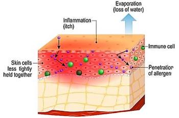
This clinical presentation is typical of atopic dermatitis, a disorder that occurs most often in infants and children. Hand dermatitis affects more than half of these patients and is frequently the only manifestation. Exacerbations and flares are very common.
Atopic dermatitis is thought to be caused by environmental factors that trigger the disease in genetically susceptible persons; it is not caused by allergens. However, consider irritant and allergic contact dermatitis in the differential. A thorough history is the most elucidating diagnostic tool and often reveals a family or personal history of atopic disorders, such as asthma or hay fever.
The major diagnostic features are:
- Severe pruritus, which can provoke an "itch, scratch, itch" cycle.
- An age-dependent distribution and morphology, which involves extensor surfaces of the extremities, face, scalp, and trunk in infants; flexural skin and neck in children; and flexural lichenification and hands and feet in adolescents and adults.
- Chronicity and recurrence of symptoms.
- A family history of asthma, allergic rhinitis, and atopic dermatitis.
Minor diagnostic features are numerous and include cheilitis, cataracts, food intolerances, elevated IgE levels, keratosis pilaris, orbital darkening, infraorbital fold, hand dermatitis, wool intolerance, hyperlinear palms, and xerosis. Three or more features from each category establish the diagnosis; the absence of pruritus makes atopic dermatitis unlikely.
Postinflammatory hypopigmented or hyperpigmented areas represent sites of previous eczematous eruptions; they are clues to past episodes of atopic dermatitis in this patient. Postinflammatory pigmentary changes occur in many chronic inflammatory disorders in which scratching or rubbing is prevalent. Typically, hyperpigmented areas are muddy brown.
The clinical presentation of atopic dermatitis is age dependent. The infantile phase features erythema, vesicles, papules, oozing, crusting, and intense pruritus. In childhood, scaly and lichenified patches and plaques that may ooze and crust are seen. The adolescent/young adult phase is characterized by dry and lichenified plaques; oozing and crusting are absent. Localized inflammation with lichenification is noted in adults. The disease often disappears in childhood or adolescence; however, the disorder continues into adulthood in up to 10% of patients.
In the acute phase only, therapy consists of compresses of Burow's solution, applied 2 or 3 times a day for 20 minutes, followed by a corticosteroid cream or ointment in a dosage appropriate for the patient's age and the severity of the eruption. Moisturizer should be applied after washing hands and bathing. Most patients respond to treatment in about 3 weeks; refractory disease in older children and adults may require ultraviolet light therapy. Antihistamines with sedative effects can be used at night to control the pruritus and to help promote sleep.
Consider 2 newer drugs--pimecrolimus and tacrolimus--for patients who cannot tolerate corticosteroids or in whom these agents are contraindicated and for those whose disease is refractory to corticosteroids. Both the macrolactam ascomycin derivative, pimecrolimus, and the macrolide immunosuppressant, tacrolimus, are indicated for short-term and intermittent long-term therapy in patients who are older than 2 years. Use pimecrolimus only in immunocompetent persons. These drugs are intended to relieve the pruritus, erythema, inflammation, and excoriation associated with atopic dermatitis.
Newsletter
Enhance your clinical practice with the Patient Care newsletter, offering the latest evidence-based guidelines, diagnostic insights, and treatment strategies for primary care physicians.


