
Here: Dr Rosen presents 5 tips about 5 skin disorders that you might not know.
Here: Dr Rosen presents 5 tips about 5 skin disorders that you might not know.
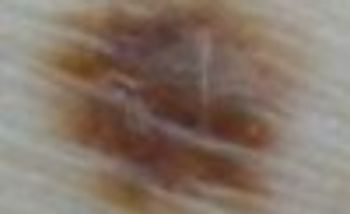
A 24-year-old woman presented with a chief complaint: “Check a funny mole.” Her boyfriend pointed out that a single mole on her back looked “different.”
Here: Ted Rosen, MD, presents 5 tips about 5 skin disorders that you might not know.
A 27 year-old woman was petting her cat, when the animal suddenly bit her on the arm. She rinsed copiously with isopropyl alcohol and applied an over-the-counter antibiotic ointment. But, 12 hours later, a large, red, swollen and exquisitely tender plaque had developed around the bite site.
Here: Ted Rosen, MD, presents 5 tips about 5 skin disorders that you might not know.

Here: Ted Rosen, MD, presents 5 tips about 5 disorders that you might not know.

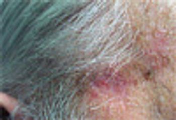
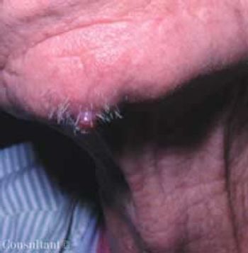
Following his routine rosacea follow-up visit, this 58-year-old man incidentally asked about a lesion on his left third digit. He pointed out a dime-sized, red, scaly patch on the dorsal aspect of the distal portion of the knuckle that extended toward the end of the finger.
While he was doing yard work, a man experienced acute, severe, burning pain.
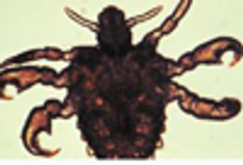
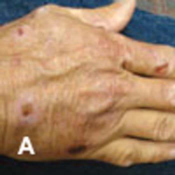
An 8-year-old child has intensely pruritic lesions on the thumbs, adjacent dorsum of the hand, soles of the feet, and navel. Vital signs are normal. The patient is otherwise healthy and has no recent history of exposure to poison ivy.
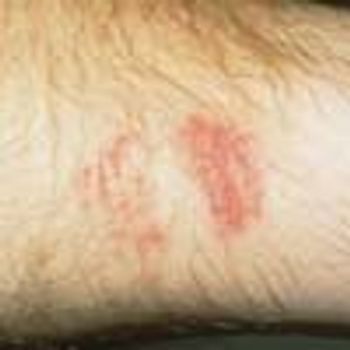
Dr Rosen invites you to match the clinical characteristics in the checklist with the disorders pictured in the photos. This month’s challenge: 2 leg lesions.

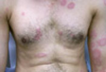
Intensely pruritic lesions of acute onset are evident on the legs of a 24-year-old woman who had no history of similar episodes. She was in excellent health and took no medications. She had spent the previous evening seated outdoors at a restaurant.
A biopsy of this asymptomatic lesion showed invasive squamous cell carcinoma without obvious perineural involvement. The lesion was excised with Mohs surgery, with good cosmetic outcome.
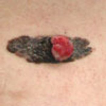
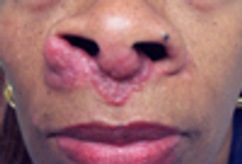
A 48-year-old Hispanic man had a tender, bleeding growth within a darkly pigmented plaque on the right flank. The pigmented lesion had been present since birth; it was previously asymptomatic. The tumor arose out of the mainly flat patch 6 months earlier and had slowly enlarged. The patient worked indoors, wore sunscreen daily, and generally avoided outdoor activities. He had no family history of skin cancer.
For several months, a 55-year-old white construction worker experienced intense burning of the skin when exposed to direct sunlight. In addition, multiple fragile blisters appeared on the dorsa of his hands and arms; these rapidly developed into crusted, superficial erosions.
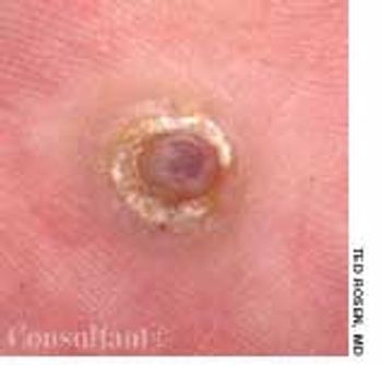
A 50-year-old woman was concerned about a nonhealing, painful lesion on the medial aspect of the left side of the nasal bridge. The lesion had been present for several weeks. The patient believed that a "cyst" had developed in the area. She had been attempting to remove it manually.
Telltale skin lesions of syphilis, gonorrhea, human papillomavirus infection, and Haemophilus ducreyi infection.

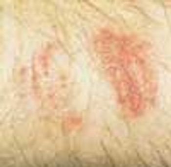
The asymptomatic skin lesions seen on this 66-year-old woman had been present for 7 months. Therapy with topical and oral antifungal agents had failed. The annular patches were pale to bright red and very slightly scaly; they affected the lower third of the patient's back and abdomen and her flanks, buttocks, and upper thighs.
A 45-year-old African American man requested treatment of “keloids” that had developed 18 months earlier. The patient also complained of dyspnea and exertion; there was no history of trauma.
A 32-year-old man who was seropositive for HIV presented with a tender lesion on his right foot of about 3 months' duration. The patient's only medication was zidovudine. His CD4+ cell count was 120/μL.
A 43-year-old man sought treatment of a “fungal nail infection” that had been present for several years. The condition had not responded to standard dosages of itraconazole, terbinafine, and fluconazole prescribed by another physician.

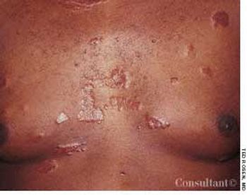
A 58-year-old man presented with a 6-month history of a mildly pruritic rash of the left axilla. The patient was in good health, took no medications, and denied any other symptoms.
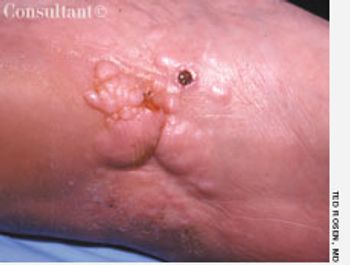
A 45-year-old woman presented with a slowly enlarging, mildly tender lesion on the left sole. The large indurated plaque was studded with multiple firm papules and nodules that involved the instep and extended onto the medial aspect of the foot. There was no regional adenopathy.
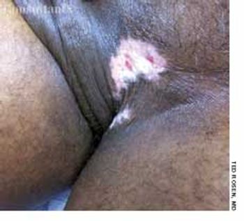
A 46-year-old man complained of “irritation” in the groin of several months' duration.
A 75-year-old Hispanic woman presented with a slowly growing, asymptomatic facial lesion of about 3 years' duration.

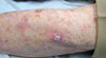
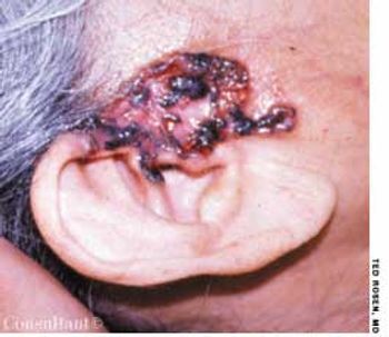
A 68-year-old man presented with a sudden-onset, 2.5 × 2-cm, rock-hard, erythematous, nontender nodule on the right side of the chest. A dense mat of telangiectases surrounded the solitary lesion. The remainder of the cutaneous examination was unremarkable.
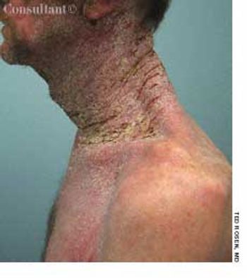
A 52-year-old white man presented with a pruritic eruption on the neck of 3 months' duration. The rash had not responded to a potent topical corticosteroid prescribed by another practitioner for the presumed diagnosis of eczema.
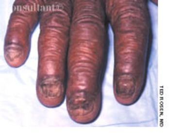
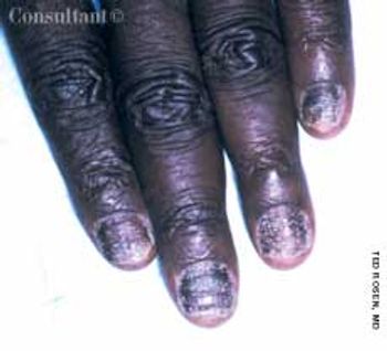
A 46-year-old man with diabetes presented for evaluation of gradual fingernail deterioration, which had failed to respond to several courses of griseofulvin and a recent 3-month course of daily terbinafine. The patient-who worked as a bartender-was otherwise healthy.
While doing yard work, a man experienced acute, severe, burning pain on relatively brief contact with the caterpillar Megalopyge opercularis. The lesion shown in the photograph developed subsequently. Each red papule represents the site of direct cutaneous envenomation by the insect's poisonous body hairs. The caterpillar can vary in color from white to dark brown, depending on the surroundings and time of year. The fuzzy hairs resemble a cat’s fur; hence the nickname “puss.”
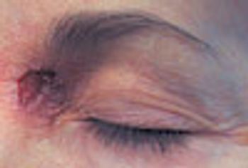
he sudden appearance of an asymptomatic, 6 x 4.5-mm, exophytic, red nodule on the chin prompted an ostensibly healthy 73-year-old nonsmoker to seek medical attention. His nodes were not enlarged, and he had no other skin lesions.
A 46-year-old man complained of “irritation” in the groin of several months’ duration. Ted Rosen, MD, of Houston noted a tender, macerated, hypopigmented plaque at the junction of the scrotum and upper inner thigh. At the periphery of the lesion was some detectable erythema and within the plaque were several small, superficial erosions.