
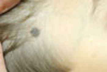
Melanoma in pre-adolescent children is extremely rare, but not totally unknown. Because this lesion had recently become larger and darker, the lesion was excised; histology confirmed a benign nevus.
Melanoma in pre-adolescent children is extremely rare, but not totally unknown. Because this lesion had recently become larger and darker, the lesion was excised; histology confirmed a benign nevus.
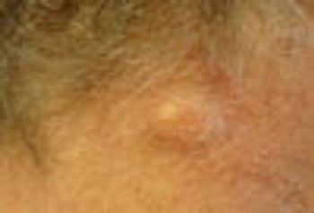
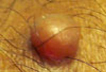
This nodule on the neck is a typical location and appearance for an epidermal cyst. If the cyst has never been inflamed or infected, it may be removable through a very small punch biopsy followed by lateral pressure or a small linear incision.
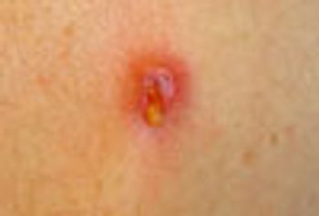
In the current milieu, the clinician should assume that MRSA is responsible for furuncles, until culture proves otherwise. Incision and drainage is the most important part of therapy, but oral antibiotics should be considered in large lesions, very young or very old patients, and when cellulitis surrounds the boil.
Given this patient's history, this wart might have been over-treated as a presumed squamous cell carcinoma had not a biopsy been obtained.
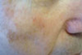
Some skin neoplasms are quite subtle and do not present as obvious exophytic lesions. This basal cell carcinoma presented as a subtle area of asymptomatic discoloration under the patient’s eye.
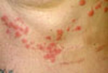
This is a classic case of herpes zoster. Note that the rash is limited to T5-6 dermatomes.
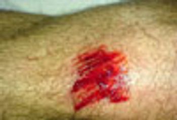
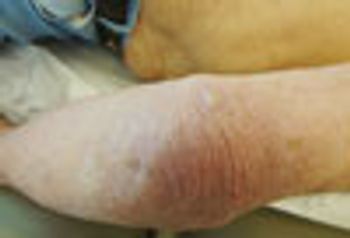
This biker asked that his wound be rinsed with hydrogen peroxide, which is now felt to be irritating to tissue and no longer recommended for use in rinsing out a traumatic wound. Water or saline is preferable. Also, superficial abrasions heal better when kept moist.
It is rare for acne of this severity to develop in normal, healthy adults. This picture strongly suggests patient manipulation of more minor lesions. When the clinical picture is unusual or improbable, always consider the possibility of factitious disease.
CT scan verified thyroid gland localization of this asymptomatic neck mass that proved to be papillary thyroid carcinoma on needle aspiration biopsy. Thyroidectomy confirmed the diagnosis of cancer.
Images: Allergic contact dermatitis, seborrheic keratosis, dermatophytosis, oro-labial herpes simplex, hereditary trichoepithelioma
This is a classic keratoderma, which can be due to a variety of congenital defects in keratinization or acquired in association with disease states. Retinoids, systemic and topical, are the treatments of choice.
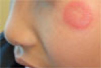
This solitary, very pruritic, annular lesion suggests dermatophytosis. KOH preparation verified the presence of Tinea faciei. This infection is usually associated with contact with a new kitten or puppy.
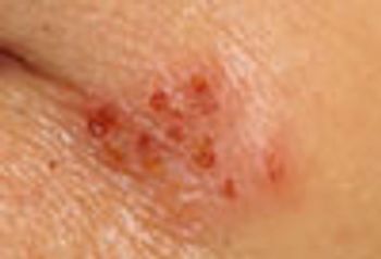
This clustered grouping of small, shallow erosions surmounting a slightly indurated plaque is characteristic for “cold sores.” Because no vesicles were present and the lesions appeared to be crusting over, the patient was advised to apply an OTC cream (docosanol 10%) per package insert instructions.
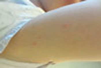
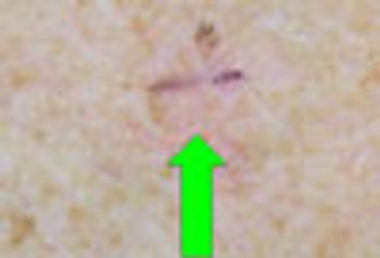
These small, pruritic papules appeared in the morning, which strongly suggests the bite of a nocturnal feeding arthropod. Insect bites arranged in clusters of 3 is typical for, but not diagnostic of, bedbug infestation.
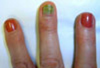
This green discoloration is the result of subungual Pseudomonas aeruginosa colonization. Pathogenic bacteria can enter this space via many causes, including trauma or underlying lesions.
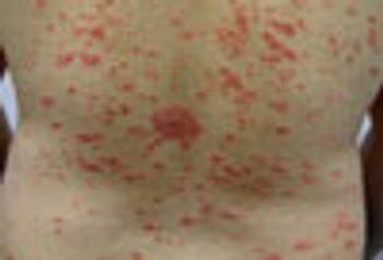
Guttate psoriasis often appears in conjunction with an upper respiratory infection, particularly streptococcal tonsillitis, as it did in this patient. This phenomenon is more common in children.
These oval, dyschromic macules surrounded by a palpable scale-like border are pathognomonic of disseminated superficial actinic porokeratosis, a disorder seen usually in those who have had considerable lifelong exposure to sunlight.
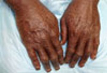
Prominent, pigmented polyangular scales on the extensor surfaces of the arms, legs, and trunk suggested recessive X-linked ichthyosis. DNA analysis demonstrated an abnormal steroid sulfatase gene, which confirmed the diagnosis.
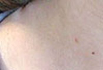
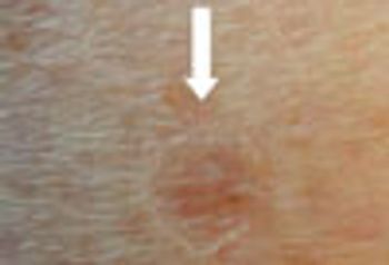
Eccentric darker pigmentation in this small asymptomatic lesion strongly suggests malignant melanoma. This patient had a history of non-melanoma skin cancer, but was unaware of the questionable growth.
This is a classic keloidal mass, which can occur in any ethic group. Because keloids can be intensely painful, they almost always require removal.
Histopathology showed this neoplasm to be a seborrheic keratosis: it might well have been a hypertrophic actinic keratosis, melanoma, or superficial basal or squamous cell carcinoma.
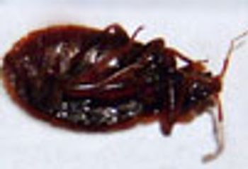
This patient wonders whether the insect he found on his headboard might be responsible for the clusters of pruritic, crusted papules on his legs. This turned out to be a bed bug.
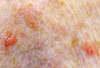
The areas that contained linear erythema, punctuated by stippled pigmentation, and a small round area of stippled pigmentation were biopsied: both showed pigmented basal cell carcinoma.
This new-onset “growth” was removed and shown to be a solitary neurofibroma, which is not an indication of any of the genodermatoses marked by a multiplicity of such neoplasms.
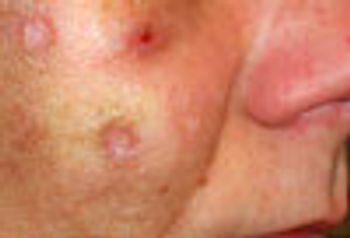
Biopsy of the 3 red “spots” in close proximity showed lichenoid keratosis-a benign lesion, probably related to seborrheic keratosis, which often mimics skin cancer.

While it might be tempting to dismiss this as a picker's nodule, lesion's thickness suggested the need for a biopsy, which revealed that the lesion was an infiltrative, invasive basal cell carcinoma. The lesson? Never hesitate to biopsy when the slightest uncertainty exists as to the correct diagnosis.
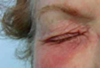
The predominantly infraorbital erythema and swelling suggested that this rash was related to a material that was being placed in the eyes and dripping from them. Patch testing showed that the patient was allergic to the preservative in the eyedrops she had been using.
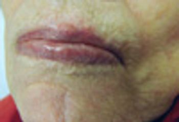
The translucent papule on the upper lip has a few small blood vessels that traverse its surface. These are morphologic features of basal cell carcinoma, a presumptive diagnosis that was confirmed by biopsy.
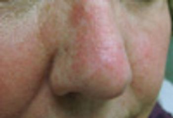
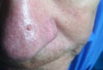
The redness on this woman's nose represents fairly severe photodamage with extensive actinic keratosis. Use of a topical therapy to achieve clearance of a field of actinic damage can be done with imiquimod, 5-fluorouracil, or ingenol mebutate creams, or diclofenac gel.
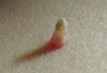
Cutaneous horns can arise on top of: seborrheic keratosis (as in this patient’s case), actinic keratoses, warts, basal and squamous cell carcinomas. Therefore, the lesion-and especially the base-must be submitted for pathologic diagnosis.