


SACRAMENTO, Calif. -- Even when HIV has all but disappeared from the peripheral blood after the start of therapy, the virus may still be lurking in the gut, slowing the restoration of immune response, according to researchers here.
HIV AIDS
Latest News

Advertisement
Advertisement
ROCKVILLE, Md. -- High-strength hydrogen peroxide, which is heavily marketed on the Internet for a diseases ranging from AIDS to emphysema, can cause serious harm or death when ingested, according to a warning issued by the FDA.
BOSTON -- The modern era of anti-HIV treatment has dramatically reduced the rate of opportunistic infections among HIV-positive children and adolescents.
VALENCIA, Spain -- With a combination of pegylated interferon and Rebetol (ribavirin), liver transplant recipients with recurrent hepatitis C (HCV) infection respond as well as non-transplants patients, researchers here reported.
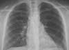
A 49-year-old man presented to theemergency department (ED) andcomplained of fever and cough thatproduced bloody sputum for 1 day.He had AIDS and recently receiveda diagnosis of large B-cell lymphoma.His most recent CD4+ cellcount was 24/µL. He had optedagainst receiving highly active antiretroviraltherapy and prophylaxisfor opportunistic infection.
Abstract: Elderly persons with active tuberculosis may present with the classic features, such as cough, hemoptysis, and fever, but some patients present with less typical signs, such as hepatosplenomegaly, liver function abnormalities, and anemia. A high index of suspicion is required when a patient presents with cough or pneumonia unresponsive to conventional therapy. Acid-fast smear and mycobacterial culture of a sputum specimen are recommended for diagnosis. For an elderly patient who tests positive with purified protein derivative, 9 months of isoniazid prophylaxis is recommended. For patients who are intolerant of isoniazid or have been exposed to or infected by an isoniazid-resistant strain, rifampin single-agent preventive therapy may be an effective alternative. (J Respir Dis. 2006;27(7):307-315)
A 37-year-old man presents with new-onset fever and abdominal pain of several days' duration. What does the PA film show, and what further action would you take to arrive at a diagnosis?
Rates of gonorrhea and chlamydial infection are highest among females 15 to 24 years old. Annual screening of all sexually active adolescents is warranted.
Early diagnosis enables patients to derive maximum benefit from highly active antiretroviral therapy (HAART). Primary care practitioners can play a key role in the timely identification of HIV infection.
ROCKVILLE, Md. - The FDA has given accelerated approval to Prezista (darunavir), a novel protease inhibitor for salvage treatment of patients with HIV.
ATLANTA - Six unrelated clusters of community-acquired methicillin-resistant Staphylococcus aureus infection involving 44 people in three states have been linked to 13 unlicensed street-corner tattoo artists.
SEATTLE - Among newly sexually active women, consistent condom use by their partners reduced the risk of human papilloma virus (HPV) infection, researchers reported.
SALT LAKE CITY - A restless leg syndrome diagnosis can be ruled out by a single question, a researcher said here.
WHITE RIVER JUNCTION, Vt ? TV and newspaper reports emerging from major medical meetings are so overstated or so lacking in context that viewers and readers would be better off paying no attention to them whatsoever, say a pair of Dartmouth investigators.
ATLANTA ? After 25 years, the AIDS pandemic has killed more than 25 million people round the world. Today, nearly 40 million people live with HIV.
A 25-year-old man reports that he has had a swollen eye for thepast several days. He noticed a small amount of yellow discharge the previousevening. He denies systemic complaints, including fever, chills, nausea, vomiting,and recent trauma. He also tells you that he has a drip in my private area.
Some sexually transmitteddiseases (STDs), such assyphilis and gonorrhea, arecenturies-old scourges; othershave attained clinicalsignificance only in recent years.Despite the availability of effectivetherapy for many of these diseases,they remain an important publichealth problem.
Patients with psychiatric disordersoften present a diagnostic challenge-even for psychiatrists. Their demeanormay not readily reveal the nature orseverity of the problem. Nevertheless,there are clues that can help you sortthrough the differential and arrive atthe correct diagnosis.
abstract: Pulmonary hypertension is an increasingly recognized complication of HIV disease. Echocardiography is the most useful imaging modality for an early diagnosis; the most frequent findings are systolic flattening of the interventricular septum, right atrial and right ventricular enlargement, and tricuspid regurgitation. Other components of the workup include comprehensive laboratory tests (complete blood cell count, measurement of prothombin time and partial thromboplastin time, hepatic profile, etc), chest radiography, pulmonary function tests with arterial blood gas analysis, ventilation-perfusion lung scanning, and spiral CT scanning. The treatment of this condition is complex and controversial, and the drug of choice has not yet been established. The therapies currently used include antiretroviral agents, bosentan, calcium channel blockers, epoprostenol, and sildenafil.
Pulmonary arterial hypertension (PAH) can be difficult to diagnose because the symptoms are nonspecific and the physical findings are usually subtle (Table). In 2004, the American College of Chest Physicians (ACCP) published clinical practice guidelines for the diagnosis and management of PAH.1 Highlights of the ACCP's recommendations for patient assessment include the following:
The authors describe a woman who presented with severe pulmonary hypertension. A cardiopulmonary cause was initially sought, but thyrotoxicosis was the underlying cause.
abstract: Pulmonary arterial hypertension (PAH) is 1 of 5 types of pulmonary hypertension (PH). Symptoms may include dyspnea on exertion, fatigue, near-syncope, and palpitations. Physical findings include lower extremity edema, jugular venous distention, and a loud P2. Findings on chest radiography, transthoracic echocardiography, and electrocardiography can suggest the presence of PAH; however, right heart catheterization is the gold standard for confirming the diagnosis and for differentiating PAH from other forms of PH. It is essential to exclude chronic thromboembolic PH, since this can be surgically corrected. The treatment of PAH depends on the severity. In addition to the standard treatments, such as diuretics and anticoagulation, more advanced treatment options include prostaglandin therapy (epoprostenol, treprostinil, and iloprost), endothelin receptor antagonists (bosentan), and phosphodiesterase inhibitors (sildenafil).
Man With Worsening Cough and Dyspnea
The mumps outbreak in midwestern states appears to be slowing, but as college students return home and engage in summer travel, it's possible that mumps will spread. Are you prepared?
Abstract: The introduction of helical CT dramatically improved the quality of CT images of the airways and other thoracic structures. Multi-detector row CT scanners have made further improvements with respect to spatial resolution, speed, and anatomic coverage. Axial CT images provide valuable information about the airway lumen and wall and adjacent mediastinal and lung structures, but they are limited in their ability to assess airway stenoses and complex airway abnormalities. These limitations can be overcome by multiplanar and 3-dimensional reconstruction images. State-of-the-art scanners allow all of the central airways to be imaged in a few seconds. This speed is particularly valuable for patients who cannot tolerate longer breath-holds and patients who may have tracheomalacia or vocal cord paralysis. (J Respir Dis. 2006;27(5):192-196)
Advertisement
Advertisement
Trending on Patient Care Online
1
Compulsive Smartphone Use Linked to Depressive Symptoms in Older Adults
2
Retatrutide Achieved Up to 22.6% Weight Loss in 2 Phase 3 Obesity Trials
3
Newer GLP-1 Therapies Linked to Fewer Alcohol-Related Hospitalizations in Adults With AUD
4
FDA Clears Roche cobas BV/CV PCR Assay for Simultaneous Vaginitis Detection
5