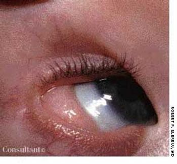
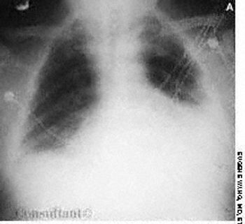
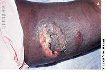
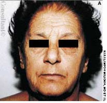
Woman With Celiac Sprue and Primary Immunodeficiency
March 2nd 2004A 34-year-old white woman presentswith a 4-month history of diarrhea,with bulky, foul-smelling stools; flatulence;diffuse abdominal discomfort;and episodic nausea and vomiting. Shehas lost 13.5 kg (30 lb) during this period.The patient has had no fever, andher medical, family, and travel historyare unremarkable.
Various Manifestations of Rheumatic Disorders: Case 6 Progressive Systemic Sclerosis
March 2nd 2004For several months, a 70-year-old woman had had dysphagia,mild dyspnea on exertion, and the Raynaud phenomenon.Her skin was waxy and edematous; 2- to 10-mm pinkishspots had appeared on her fingers, palms, and oral mucousmembrane over the past 2 weeks. These disappearedcompletely with pressure. Subcutaneous calcific depositswere present on the extensor surfaces of the forearms.
Various Manifestations of Rheumatic Disorders: Case 5 Rheumatoid Nodules
March 2nd 2004A 65-year-old woman, who was confined to a wheelchairbecause of severe rheumatoid arthritis, was concernedabout nodules that had erupted on her fingers and handsduring the previous 3 weeks (A). Her medical historyincluded colon cancer, chronic renal insufficiency, anemia,and hypertension. The nonpruritic nodules were painfulwhen they began to form under the skin; however, oncethey erupted, the pain disappeared.
Various Manifestations of Rheumatic Disorders: Case 3 Rheumatoid Vasculitis
March 2nd 2004Ten weeks before presentation, this55-year-old woman noticed decreasedsensation in her feet and a bluish discolorationof her toes. These symptomsprogressed rapidly, and pain andcoldness in both feet increased in intensity.Her feet subsequently becamegangrenous. Her seropositive arthritishad been diagnosed about 6 yearsearlier. The disease had been wellcontrolled until about 10 weeks beforethis photograph was taken.
Various Manifestations of Rheumatic Disorders: Case 2 Heberden Nodes
March 2nd 2004Bilateral swelling and pain in the distal interphalangeal (DIP) joints for severalmonths brought this 65-year-old woman to her physician. She complained alsoof stiffness in the region of the DIP joints when she arose in the morning andafter short periods (less than 15 minutes) of inactivity. A history such as this,in conjunction with the appearance of the patient’s hand, is typical of Heberdennodes, which are a manifestation of osteoarthritis (OA).
Tinea Types: Common Dermatophyte Infections Case 8 Tinea Capitis
February 1st 2004The parents of a 3-year-old girl sought evaluation of their daughter’s hair loss.During the past several months, a large patch of alopecia with scaling had developed.The differential diagnosis included seborrhea, trichotillomania, andtinea capitis.
The 10 Most Common Prescribing Errors: Tips on Avoiding the Pitfalls
February 1st 2004Numerous factors contribute to the medication errors that kill up to 98,000 patients each year. Unnecessarily high dosages can result in increased side effects with only a small therapeutic benefit, especially in elderly patients. Lack of patient information-such as a history of allergies or adverse drug reactions-is another cause of error and injury. Communication failures include the use of ambiguous abbreviations, misinterpretation of verbal orders, and lack of timely response to a patient's medication-related symptoms. Dosing errors are common in children because of variability in dosage expressions in drug references. Remedies for prescribing errors are described in detail here.
Fibromyalgia Syndrome: Can It Be Treated?
February 1st 2004Treatment of fibromyalgia syndrome (FMS) is a challenge. However, most patients benefit from appropriate management. Essential to treatment are a physician's positive and empathetic attitude, continuous psychological support, patient education, patience, and a willingness to guide patients to do their part in management. Other important aspects involve addressing aggravating factors (eg, poor sleep, physical deconditioning, emotional distress) and employing various nonpharmacologic modalities (eg, regular physical exercise) and pharmacologic therapies. Drug treatment includes use of tricyclic medications alone or in combination with a selective serotonin reuptake inhibitor, and other centrally acting medications. Tender point injection is useful. It is important to individualize treatment. Management of FMS is both a science and an art.
Is There a Medical Explanation?
February 1st 2004A 13-year-old girl of African American descent is brought to the pediatrician’s office becauseof a lesion on her neck. The girl’s mother had telephoned the office before the visit, statingthat the lesion resembled a blister at first, but now looked like a burn.
Images of Malignancy: Case 4 Peritoneal Carcinomatosis
January 2nd 2004Vague abdominal pain, malaise, anorexia,and the loss of 10 lb in 2months prompted a 65-year-old manto seek medical evaluation. A yearearlier he had undergone surgery forstage III carcinoma of the sigmoidcolon. Because metastases to thelymph nodes were found in the resectedcolon, the patient was given postoperativechemotherapy. Histologicexamination revealed poorly differentiatedadenocarcinoma.
Images of Malignancy: Case 2 Papillary Carcinoma of the Thyroid
January 2nd 2004A 40-year-old man was concerned about an enlarging painlessmass on the right side of his neck that had been presentfor 6 months (A). The patient reported no other healthproblems; his medical history was unremarkable, and hewas taking no medications.There was no family or personal history of thyroiddisease or of exposure to radiation. Thyroid function testresults were within normal limits. A chest film revealed nopathology.
Eye Signs of Systemic Disease: Case 5 Central Retinal Vein Occlusion
January 1st 2004An 87-year-old woman complained ofseeing a red tinge on the wallpaper inher house through her right eye. Thepatient had mild memory loss andmoderate hypertension, for whichshe took atenolol. She had quit smokingcigarettes many years earlier.
Eye Signs of Systemic Disease: Case 6 Diabetic Maculopathy
January 1st 2004During an annual eye examination, a 65-year-old womanwith a 5-year history of type 2 insulin-dependent diabetescomplained that her vision had slightly worsened in botheyes. Her best corrected visual acuity was 20/30 in botheyes.