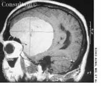
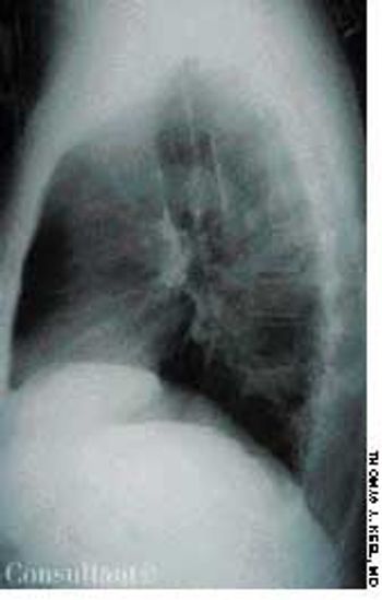
Evaluation of intermittently discolored, cold fingers was sought by a 39-year-old woman with long-standing anorexia nervosa. The patient had never smoked and was not taking any vasoconstrictive drugs.

Evaluation of intermittently discolored, cold fingers was sought by a 39-year-old woman with long-standing anorexia nervosa. The patient had never smoked and was not taking any vasoconstrictive drugs.

A 45-year-old man complained of blood in his urine. The patient had a 7-year history of chronic renal failure secondary to hypertension; he had undergone hemodialysis for the past 5 years.

An 83-year-old woman-who had not visited a physician for 20 years-presented to the emergency department with a 1-day history of urinary retention and a 1-month history of gross vaginal bleeding. The mass on the left side of her face was a secondary finding.

A 15-year-old boy presented with sharp, right lower quadrant abdominal pain, nausea, vomiting, and fever of 2 days' duration. His white blood cell count was 15,000/mL with a shift to the left on the differential. Right lower quadrant rebound tenderness was noted.

A 65-year-old man had had an asymptomatic lesion on his left lateral lower leg for several weeks. The dark maroon, almost black, 3- to 4-mm, circular, elevated lesion had a convoluted surface of dilated vessels.

A 35-year-old woman noticed that her right upper eyelid started to droop as the day progressed. She denied other ocular problems, including decreased visual acuity, pain, or diplopia. The patient had no generalized fatigue, difficulty in swallowing, or weakness of her arms or legs.

After this 9-year-old boy complained of “growing too fast” during the past 1 to 2 years, his parents sought medical advice. He had no significant past medical history.

A 63-year-old African American man presented with orthopnea and pedal edema that had worsened during the past 4 months. Macroglossia was noted.

The diagnosis of acute appendicitis is usually straightforward. When the presenting symptoms are atypical, abdominal ultrasonography can be of diagnostic assistance if it shows the thickened walls of the appendix and a distended, noncompressible lumen.

A 56-year-old man who consumed moderate amounts of alcohol was awakened by an intense burning pain in the right great toe; local erythema and edema were also present. Within hours, the pain became excruciating, and the same symptoms developed in the left great toe. Acetaminophen provided no relief. The patient's serum uric acid level was 8.8 mg/dL.

Three days after having eaten fish, a 66-year-old woman with a known allergy to fish and a history of schizophrenia was brought to the emergency department because of macroglossia-a presentation of anaphylaxis. The patient refused airway management (intubation or cricothyrotomy) and was therefore admitted to the medical intensive care unit for monitoring of her airway and hemodynamic status. She received corticosteroids, ranitidine, diphenhydramine, epinephrine, and oxygen (via nasal cannula).

Three years ago, the young man pictured here was told that he might have acromegaly, but he ignored this warning. Now, at age 32, he consulted an endocrinologist because he realized his facial features and voice were becoming increasingly coarse. Soft-tissue swelling of his hands was also noted.

This photo was taken minutes after the birth of a female newborn and reveals a purple discoloration of the right buttock.

The patient reported that he had noticed the facial eruption a couple of months earlier. On examination, the lesion appeared more plaque-like and dense than is typical of rosacea. The differential diagnosis included lupus, mycosis fungoides, rosacea, and angiosarcoma.

These photos are of the left hand of a full-term, newborn infant with focal ring constrictions of the third, fourth, and fifth fingers; secondary edema and discoloration are also seen.
A 5-day history of pain and swelling in the right third finger (A) were the complaints of a 76-year-old man. A few days earlier, another physician had prescribed indomethacin, 25 mg tid, but it had not helped, and the patient believed that his condition had worsened. He had had an attack of gout 5 years before but had not been taking any maintenance medication. The distal interphalangeal (DIP) joint of the affected finger was now erythematous and tender, with chalky subcutaneous deposits. A diagnosis of acute gouty arthritis and gouty tophus was made.

A 51-year-old woman complained of mild pain in the Achilles tendon area of both feet. This was aggravated when she stood for prolonged periods and when she wore tight, high-heeled shoes. Physical examination results were unremarkable except for a disputable palpable thickness of the Achilles tendons, especially on the right foot.

A 70-year-old man was brought from a nursing home to the emergency department with abdominal distention and vomiting of recent onset and a 2-day history of fever and abdominal pain. The patient had chronic obstructive pulmonary disease, type 2 diabetes mellitus, and hypertension. His gastric feeding tube, which had been placed via percutaneous endoscopic gastrostomy, was blocked.

Cyanosis involving the hands and toes of a 12-year-old girl was first noted 3 months before medical evaluation was sought. Her fingers and palms were red, the nail beds were cyanotic, and her palms were cold and sweaty. The girl's toes were cyanotic when she was standing. Her hands and feet reverted to a normal color when the limbs were elevated.

Systemic lupus erythematosus (SLE) was diagnosed in an 18-year-old man who presented with polyarthritis, fever, hypoxia, fatigue, anemia, neutropenia, and abnormal urinary sediment. A renal biopsy showed diffuse mesangial proliferative glomerulonephritis (World Health Organization class II). Serologic tests were positive for fluorescent antinuclear antibody (FANA), SS-A, SS-B, anti-Sm and anti-dsDNA antibodies, and rheumatoid factor; a direct Coombs' test result was positive as well.