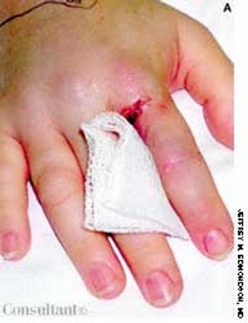
This condition, which accounts for about 30% of cases of intestinal obstruction among neonates, is characterized by the inspissation of thick, tenacious meconium in the bowel. The most common cause is cystic fibrosis; approximately 6% to 20% of infants with cystic fibrosis have meconium ileus. Hyperviscous mucus secreted by abnormal intestinal glands, an abnormal concentrating process in the proximal small intestine, and a deficiency of pancreatic enzymes have been implicated in the pathogenesis. The histologic hallmark is distention of the goblet cells in the intestinal mucosa.
All News
For 2 months, a 31-year-old woman had had dyspnea and dull, continuous retrosternal pain. She was admitted to the hospital, and a helical CT scan of the thorax identified a saddle pulmonary embolism. An ultrasonogram revealed deep venous thrombosis (DVT) in the left leg. Intravenous heparin was given; the patient was discharged, and warfarin was prescribed.

A 40-year-old man was hospitalized with the superficial, reddening, and painful facial lesion seen here. Its borders were sharp, and it had developed rapidly. The patient had a temperature of 40°C (104°F) and chills. His erythrocyte sedimentation rate was elevated, and he had neutrophilic leukocytosis.

Headache and vomiting suddenly developed in a 41-year-old woman who was 16 weeks pregnant. The next day, she suffered an episode of tonic-clonic seizures associated with a 15-minute loss of consciousness.

A 35-year-old man, a sea and white-water kayaking instructor, complained of pain and congestion in the right ear. Otoscopy revealed bilateral external auditory canal stenosis with impacted cerumen and otitis externa on the right.

A 68-year-old man presented with a sudden-onset, 2.5 × 2-cm, rock-hard, erythematous, nontender nodule on the right side of the chest. A dense mat of telangiectases surrounded the solitary lesion. The remainder of the cutaneous examination was unremarkable.

While on the job, a 24-year-old construction worker accidentally shot a nail through his right index finger with a nail gun. Roentgenographic studies revealed no damage to the bone. No neurovascular deficit was noted on examination.
Tortuous, dilated varicosities; multiple smaller caliber abnormal perforating vessels; and chronic brawny edema of chronic venous insufficiency (CVI) were seen on a 70-year-old man's left leg. He reported that the edema and discoloration had worsened over the last 15 years. The brawny edema stopped just above the ankle, indicating that compression by the patient's sock controlled the signs and symptoms of CVI.

After suffering with a severe, disabling headache for 2 weeks, a 20-year-old soldier sought medical treatment. He had no significant medical history other than his 6-year history of smoking.

A 22-year-old man complained of progressive shortness of breath and abdominal distention. Three years before, he had completed chemotherapy for Hodgkin's disease and had since been in remission. Recently, he had been treated for tonsillitis with oral antibiotics.

For 2 years, a 40-year-old oil-field worker wore heavy, steel-toed work boots all day. He experienced weeping and white discoloration at the center of both distal soles. At night, with his boots off, the feet would dry and hurt. There was no pruritus.
An 82-year-old man complained of dry, gritty eyes with chronic, bilateral tearing. He stated that friends tell him his eyes are always red.

A 42-year-old woman complained of facial puffiness, fullness, and redness for 2 weeks, which were gradually becoming more severe. She also noticed a sensation of “heaviness” in her head. The patient had no significant past medical history. She denied having a cough, shortness of breath, hoarseness, allergies of any kind, and neurologic deficits. Her weight and appetite were unchanged. She had smoked a pack of cigarettes every day for the past 20 years.
Cutaneous photosensitivity followed by acute, then chronic, skin lesions on sun-exposed areas characterizes this disorder.

Two 7-year-olds show the purpuric rash of the lower body and legs that is typical of Henoch-Schönlein purpura. This disease is a vasculitis that chiefly affects small vessels of the skin, joints, gastrointestinal tract, and kidney.
A 55-year-old woman, who had fairly well-controlled type 2 diabetes for 15 years, noticed a slow, progressive decrease in the visual acuity of her right eye during the past 6 to 8 weeks.

A 72-year-old woman who was being evaluated for medical clearance for an elective cholecystectomy was asymptomatic, except for biliary colic. She had had pulmonary tuberculosis (TB) 40 years earlier, for which she had undergone surgery on the right hemithorax-a fact borne out by a thoracotomy scar. An x-ray film of her chest, shown here, demonstrates numerous plastic balls in the right thoracic cavity. This is an example of collapse therapy for pulmonary TB, which was practiced years ago, before the availability of effective antituberculous medications.

A 3-mm long, double-tipped, polypoid lesion appeared just anterior to the anus on an 8-month-old girl 2 days earlier. The lesion was excised in the office: a local anesthetic was administered, and the base was lightly electrodesiccated. Antibiotic ointment was applied until the area healed. Pathologic findings identified an infantile perianal pyramidal protrusion.
During a routine preschool examination, it was noted that this 4-year-old boy, who weighed 30.8 kg (68 lb), did not seem to rotate his arms. The child's mother stated that she first noticed this 2 or 3 years earlier but thought it was related to her son's obesity. The youngster appears to function normally; he is able to write and play football and basketball. No other family members are known to be similarly affected.

For 1 week, a 22-month-old boy experienced gradually increasing facial swelling followed by swelling of the abdomen and legs. His mother reported that the child's urinary output had markedly decreased. During this period, the youngster gained 5 lb.