
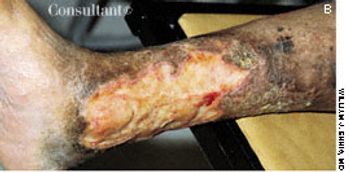
This 64-year-old woman has had rheumatoid arthritis for 15 years; her hands show classic rheumatic changes. She also had a leg ulcer that had failed to heal in 8 years despite three skin grafts.
This 64-year-old woman has had rheumatoid arthritis for 15 years; her hands show classic rheumatic changes. She also had a leg ulcer that had failed to heal in 8 years despite three skin grafts.

Various types of cutaneous lesions may occur in association with RA, including rheumatoid nodules, rheumatoid neutrophilic dermatitis, vasculitis, palpable purpura, and pyoderma gangrenosum. Many of these manifestations-including rheumatoid nodules-are specific for RA. The presence of these nodules is associated with seropositive disease and with a more severe, erosive clinical presentation. The nodules appear in 20% to 30% of patients with RA. Sites of predilection are those subject to shear stress, including the subcutaneous tissues over the extensor aspects of the elbow region, over the sacrum in bedridden persons, and at the pericardial and pleural surfaces.

This 60-year-old man has had rheumatoid arthritis (RA) for 40 years. Typical of progressive, long-standing disease is the deformity seen here in his hands and feet.

Cutaneous and subcutaneous lesions of the extremities may be clues to the presence of rheumatic diseases such as systemic lupus erythematosus (SLE), rheumatoid arthritis (RA), and the vasculitides. The history and physical examination can generally help confirm the cause. Skin biopsy is sometimes necessary for a definitive diagnosis; useful results depend on a technique that gives the depth necessary to see the pathology and proper interpretation of biopsy specimens by an experienced dermatopathologist.

This 20-month-old girl was born to a 28-year-old mother at 38 weeks' gestation. The pregnancy was uncomplicated, and vaginal delivery was normal. The infant sat with support at 10 months of age, sat without support at 12 months, crawled at 13 months, and walked at 18 months. She had not yet begun to talk at 20 months. The child was noted to have frequent laughing episodes and often made flapping movements with her hands.
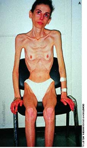
A 30-year-old woman was brought to the hospital with syncope, bradycardia, and hypotension. For the past 6 years, she had vomited after eating meals and after occasional episodes of binge eating.
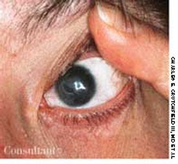
Severely limited vision plagues this 27-year-old woman with aniridia, the bilateral absence of the iris of the eye.

This x-ray film of a man in his 50s illustrates an extreme case of renal osteodystrophy. On examination, the patient's legs were very tender and one could actually see the femur bend in the examiner's hands as if it had a greenstick fracture.
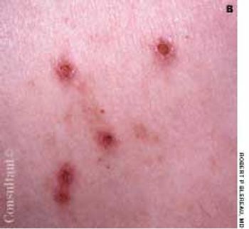
Highly pruritic, 2- to 4-mm, papular lesions with central ulceration erupted on the back of a 66-year-old woman. She had had 2 similar outbreaks in the past. The patient was taking conjugated estrogens, alprazolam, and alendronate.
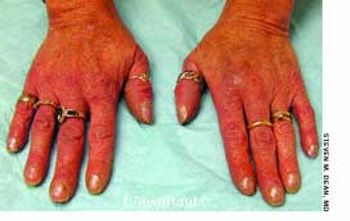
Evaluation of intermittently discolored, cold fingers was sought by a 39-year-old woman with long-standing anorexia nervosa. The patient had never smoked and was not taking any vasoconstrictive drugs.
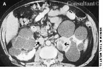
A 45-year-old man complained of blood in his urine. The patient had a 7-year history of chronic renal failure secondary to hypertension; he had undergone hemodialysis for the past 5 years.
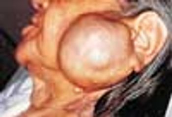
An 83-year-old woman-who had not visited a physician for 20 years-presented to the emergency department with a 1-day history of urinary retention and a 1-month history of gross vaginal bleeding. The mass on the left side of her face was a secondary finding.
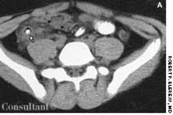
A 15-year-old boy presented with sharp, right lower quadrant abdominal pain, nausea, vomiting, and fever of 2 days' duration. His white blood cell count was 15,000/mL with a shift to the left on the differential. Right lower quadrant rebound tenderness was noted.
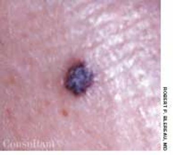
A 65-year-old man had had an asymptomatic lesion on his left lateral lower leg for several weeks. The dark maroon, almost black, 3- to 4-mm, circular, elevated lesion had a convoluted surface of dilated vessels.
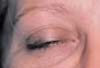
A 35-year-old woman noticed that her right upper eyelid started to droop as the day progressed. She denied other ocular problems, including decreased visual acuity, pain, or diplopia. The patient had no generalized fatigue, difficulty in swallowing, or weakness of her arms or legs.

After this 9-year-old boy complained of “growing too fast” during the past 1 to 2 years, his parents sought medical advice. He had no significant past medical history.
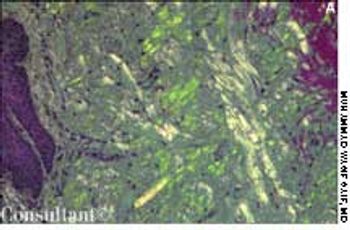
A 63-year-old African American man presented with orthopnea and pedal edema that had worsened during the past 4 months. Macroglossia was noted.
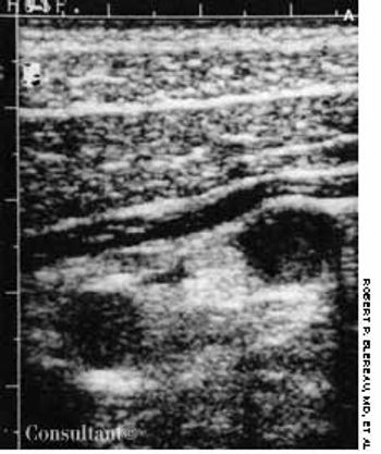
The diagnosis of acute appendicitis is usually straightforward. When the presenting symptoms are atypical, abdominal ultrasonography can be of diagnostic assistance if it shows the thickened walls of the appendix and a distended, noncompressible lumen.
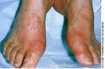
A 56-year-old man who consumed moderate amounts of alcohol was awakened by an intense burning pain in the right great toe; local erythema and edema were also present. Within hours, the pain became excruciating, and the same symptoms developed in the left great toe. Acetaminophen provided no relief. The patient's serum uric acid level was 8.8 mg/dL.
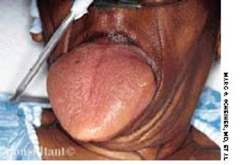
Three days after having eaten fish, a 66-year-old woman with a known allergy to fish and a history of schizophrenia was brought to the emergency department because of macroglossia-a presentation of anaphylaxis. The patient refused airway management (intubation or cricothyrotomy) and was therefore admitted to the medical intensive care unit for monitoring of her airway and hemodynamic status. She received corticosteroids, ranitidine, diphenhydramine, epinephrine, and oxygen (via nasal cannula).