
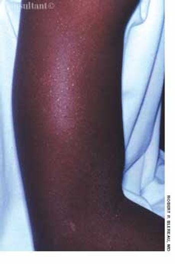
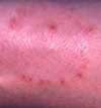
For 2 months, an asymptomatic rash had been present on the upper arms of a 16-year-old boy of normal weight. The rash, as seen on the patient's right arm, consisted of abundant fine papules. He had no other lesions.
For 2 months, an asymptomatic rash had been present on the upper arms of a 16-year-old boy of normal weight. The rash, as seen on the patient's right arm, consisted of abundant fine papules. He had no other lesions.
A 5-month-old boy's head hit the floor after he tumbled from his mother's arms. No injury was noted by the parents, and the infant immediately began to cry. His mother brought him to the emergency department just to be sure he was not seriously injured.
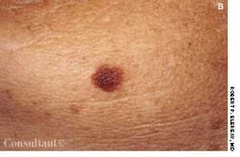
This asymptomatic pigmented lesion on the left posterior jaw of a 52-year-old man had been present for 6 years, with no evidence of recent change. At the patient's request, the lesion was excised.
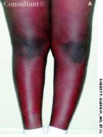
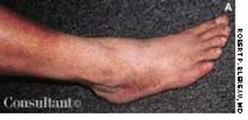
A 16-year-old African American boy complained of exertional pain below his left knee that severely limited his ability to participate in sports. The patient had had bilateral bowed legs until his early school years, when the right knee straightened. For the past year, exertional pain had been present below the left knee in the epiphyseal area.
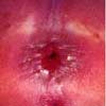
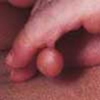
A 3-mm long, double-tipped, polypoid lesion appeared just anterior to the anus on an 8-month-old girl 2 days earlier. The lesion was excised in the office: a local anesthetic was administered, and the base was lightly electrodesiccated. Antibiotic ointment was applied until the area healed. Pathologic findings identified an infantile perianal pyramidal protrusion.
A 49-year-old man was concerned about a right flexor forearm lesion that had been increasing in size for 6 weeks. The light pink, well-demarcated, 5-cm, circular lesion featured slight peripheral elevation with ulceration, crusting, and a relatively clear central area. A culture of material from the lesion was negative for fungi. A potassium hydroxide evaluation was not performed.
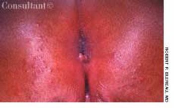
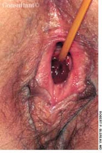
An excrescence at the 6 o'clock position at the anal verge is evident on this 8-day-old girl's anus. A hemorrhoid needs to be considered in the differential diagnosis of rectal bleeding in all age groups, since the condition can be present in neonates as well as adults.
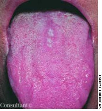
A 35-year-old woman complained of pain or a burning sensation on her tongue when she ingested hot or acidic food and beverages. The white patch had spontaneously developed on her tongue 3 months earlier.
A 62-year-old man consulted his physician hoping to confirm his suspicion that this lesion was benign and caused by skin friction from his watch. The 0.75-cm, purplish red, raised, rounded, dome-shaped mass with a keratin-filled crater at its top was on the patient's left distal extensor forearm. The lesion had developed within 3 weeks.
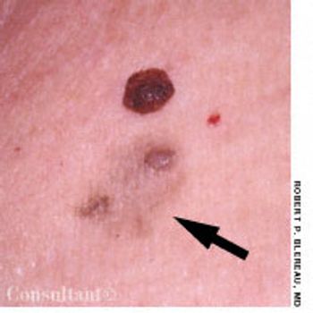
Two asymptomatic lesions that had been present for 1 year on the left upper chest of a 61-year-old man were excised. Pathologic examination revealed the raised, dark brown medial lesion to be an interdermal nevus; the lateral, light tan lesion with papules (arrow) was a papillomatous interdermal nevus with primary macular amyloidosis.

A 71-year-old woman was brought to the hospital with blunt abdominal trauma suffered in an automobile accident. She complained of pain in the chest and abdomen.
Following a cholecystectomy, an indwelling urethral catheter was placed in a 51-year-old woman with urinary retention. Five days later, the patient complained of a burning sensation at the site. A rounded, swollen, hemorrhagic area surrounding the catheter was noted, and a urinary tract infection was diagnosed.
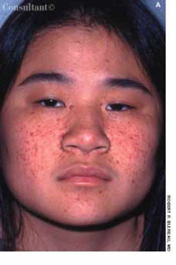
Tan-pink acneiform lesions on the face of a 15-year-old girl had not responded to topical acne therapy. A 1 × 0.5-cm, elevated subcutaneous nodule was noted on the right lateral knee. The lesions on her face and knee had been present for 11 years. The family history was noncontributory.
Following an uneventful pregnancy, a 25-year-old woman was delivered of an infant girl by repeat cesarean birth with occiput anterior presentation at 39 weeks gestation. The deformity was readily evident at birth.

The pigmented hairy nevus that covers the entire right upper arm of this 18-month-old boy has been present since birth. More than 95% of large, congenital pigmented nevi have a hairy component.
A 76-year-old man reported a 3-month history of an asymptomatic, raised, reddened lesion on his penis. The patient had type 2 diabetes mellitus. In 1994, a basal cell carcinoma had been excised from his chest and, 3 years later, a squamous cell carcinoma was excised from his left temple.
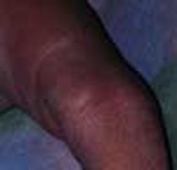
A 56-year-old man had an asymmetric, maculopapular, sharply demarcated, pruritic, excoriated dermatitis on his upper thighs. The eruption had been present for 2 to 3 weeks.
Tiny, asymptomatic "bumps" had been present for 4 years on the posterolateral surface of the upper arms of a 34-year-old woman. Similar but less severe eruptions also appeared on her forearms and anterior thighs.
The raised, fleshy lesion over the lateral conjunctiva of this 7-month-old girl's right eye has been present and gradually enlarging since birth. Consultation with a pediatric ophthalmologist yielded a diagnosis of lipodermoid choristoma, a dermoid-type congenital malformation of the conjunctiva or epibulbar region. Histologic examination reveals benign fatty and ectopic lacrimal tissue and epidermal structures.
This neonate was born with an extra hypoplastic digit attached to the lateral aspect of the left little finger's middle phalanx. When informed of this anomaly on her daughter's hand, the mother registered neither shock nor surprise. Mother, grandmother, and other family members were similarly affected.
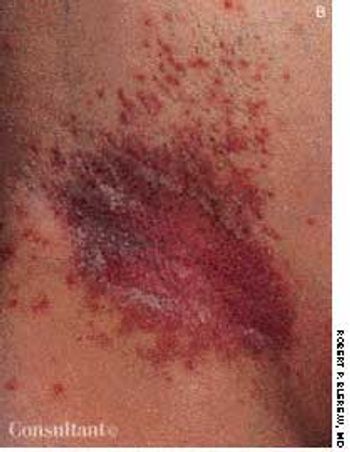
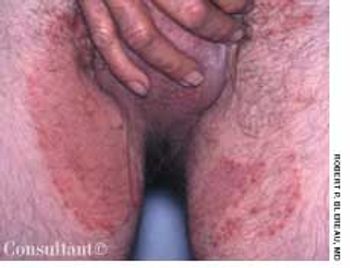
The erythematous rash with punctate satellite lesions seen on the axillary and inframammary areas of a 46-year-old woman are typical of the lesions of moniliasis, or candidiasis. The patient reported that the pruritic rash had erupted 2 months earlier and worsened during the summer heat.
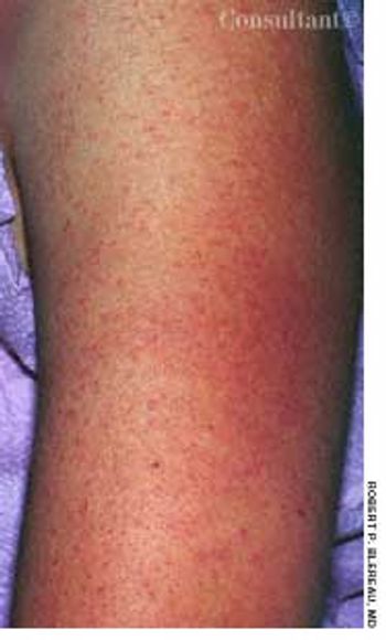
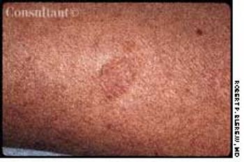
These orange-to-brown macules with red puncta, or cayenne pepper spots, are typical of Schamberg's disease (progressive pigmented purpuric dermatosis). The cause of this disorder is unknown, but it may be related to a cellular immune reaction or drug reaction.
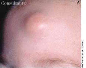
A 0.5-cm mass with overlying erythema on the forehead of a 5-month-old girl was first noticed after she had been hit on the head with a rattle. Excision of the mass was deferred because it was considered to be a hemangioma.
These pruritic but otherwise asymptomatic lesions on the right upper arm of a 77-year-old woman first appeared about 1 year before she sought medical consultation. The patient's history included frequent, generalized pruritus, which was believed to be secondary to long-standing type 1 diabetes mellitus.
Concern about this flat, tan, atrophic, well-marginated, dime-sized lesion prompted a 44-year-old woman to seek medical advice. The lesion, which was mildly pruritic, had appeared 2 months earlier.
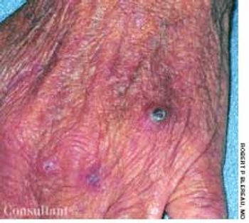
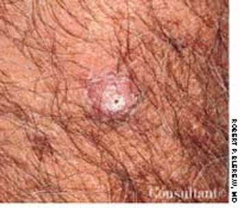
For 1 month, a 66-year-old man had had an asymptomatic lesion on the dorsum of his left hand. The flesh-colored, dome-shaped, maroon-crusted lesion measured 0.7 cm in diameter and was located over the fourth knuckle. The patient had chronic obstructive pulmonary disease but was otherwise in good health. He was seronegative for HIV.
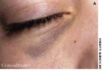
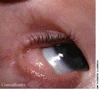
The mother of this 10-year-old boy noticed the shiner on her son's right eye after he awakened one morning. The child had bronchitis and a persistent cough for the past few days. Particularly alarmed by the extensive subconjunctival hemorrhage that appeared the next morning, the parent brought her child for medical evaluation. The youngster had no history of injury.
The bilateral breast hypertrophy seen on a 12-day-old boy's chest began 4 days earlier. This anomaly results from elevated levels of maternal circulating estrogen in late gestation that crosses the placenta and stimulates the baby's breasts. Both sexes are affected equally.
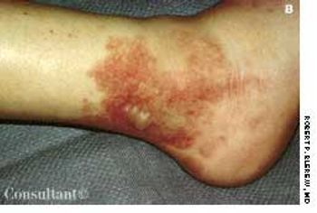
A 16-year-old girl was bothered by ankle pain and “red spots” on her lower legs. These symptoms cleared in a few days without treatment. Six weeks later, after returning from an all-day outing at a fair, she noticed that the spots reappeared and hemorrhagic lesions developed on the right ankle and left heel. After removing her shoes, the teen-ager felt severe pain in both ankles, particularly the right.
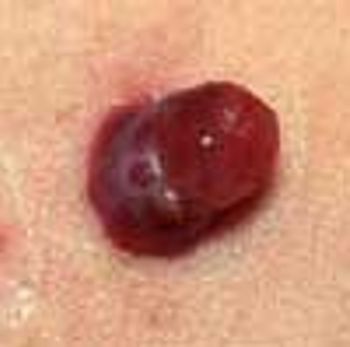
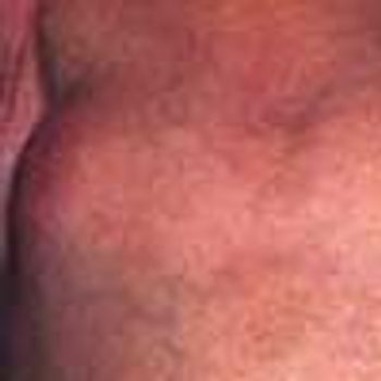
A 1 × 0.5-cm hemorrhagic, polypoid lesion that had been present for 2 months on a 13-year-old boy's left anterior chest was excised. Pathologic examination confirmed the diagnosis of pyogenic granuloma.