
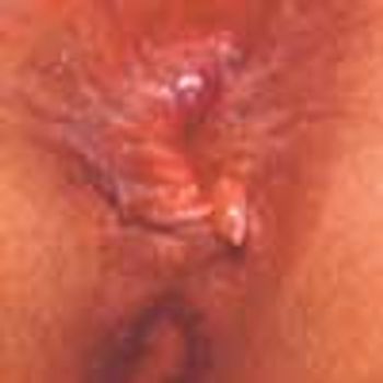
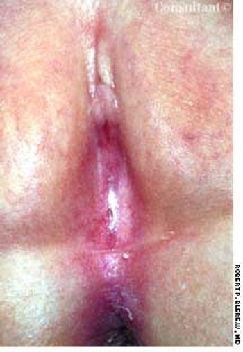
The lesion is a hypertrophied papilla--an enlarged teat-like structure that may develop in an anal crypt. Treatment options include surgical excision, electrodesiccation, or cryotherapy.
The lesion is a hypertrophied papilla--an enlarged teat-like structure that may develop in an anal crypt. Treatment options include surgical excision, electrodesiccation, or cryotherapy.
A medium-sized brown pigmented lesion had been present on the right temple of a 61-year-old man since birth. The patient complained that during the last several months the lesion had formed a fine scale and begun to itch. Actinic keratoses and inclusion cysts were also noted on his face.
During week 28 of her pregnancy, a 28-year-old woman presented with a gingival hemangioma between the right upper central incisor and canine tooth. The asymptomatic lesion arose during the thirteenth week of her pregnancy; it gradually enlarged and bled occasionally with minor trauma.
The lesion on this 6-year-old boy occupies almost the entire left side of his nose. The mother attributed it to an injury her son had sustained 2½ weeks earlier, when he was hit in the face by a baseball.
A 7-year-old girl presents for evaluation of a pink, polypoid, 2 x 2-mm lesionon her inner lower lip, which has been present for several weeks. The child isasymptomatic and denies any trauma to the mouth.
A 1 X 0.5-cm hemorrhagic, polypoid lesion that had been present for 2 monthson a 13-year-old boy’s left anterior chest was excised. Pathologic examinationconfirmed the diagnosis of pyogenic granuloma.
An 80-year-old man has had an asymptomatic, flesh-colored swelling on his right ear for 4 to 5 months. In the center is a 1-mm white scab pointing downward from the helix. At times, the patient shaves a white spicule that grows in this crusted area. He sleeps on his right side and does not use a cell phone.
For 3 years, a 23-year-old man has had this asymptomatic, 0.75-cm, polypoid lesion on his left wrist.
Telltale skin lesions of syphilis, gonorrhea, human papillomavirus infection, and Haemophilus ducreyi infection.
These pinpoint pustules, some with excoriations, and surrounding erythema appeared on the posterior trunk and outer arms of a 15-year-old boy after he had wrapped his upper body in a wool blanket. These lesions were occasionally pruritic, especially on the arms, where most of the excoriations were noted.

A 41-year-old man fell 3 ft into a bilge; he landed on his left leg and experienced immediate generalized pain in that knee. Three days later, he consulted his physician, who found minimal effusion in the knee and tenderness of the medial collateral ligament (MCL). No abnormalities were seen on plain x-ray films.
The mother of a 1-year-old girl observed that for 1 week her child seemed to be uncomfortable during the night and when she urinated.

After being hit on the head during a football game, a 16-year-old experienced several seconds of complete vision loss in the left eye. A few days later, he noticed the onset of blurred vision in the same eye, which progressively worsened over several weeks.
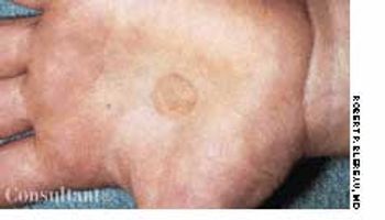
Identical circular, chocolate-colored spots developed on a 4-year-old boy's palms 2 weeks ago, according to the child's mother. The sharply demarcated, macular lesions were asymptomatic.
Encountered rarely these days, scarlet fever is believed to be caused by sensitization to an erythrogenic toxin produced by strains of group A β-hemolytic streptococci. Thus, previous exposure to the toxin is necessary for development of the rash seen here-fine, sandpaper-like, and papular on an erythematous background. It usually begins on the trunk and spreads over the entire body within hours or days. Scarlet fever is unusual in infancy, possibly because of maternal transfer of antibodies.
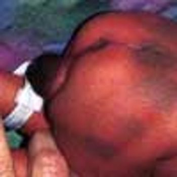
Large, blue-black, macular areas over the buttocks and presacral regions were present at birth in this black child. Significant hyperpigmentation of the genitals also was evident. Scrotal hyperpigmentation is not an uncommon finding in a black newborn. However, the intensity of the penile hyperpigmentation in this baby is unusual.
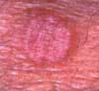
A new lesion recently arose on the right flexor forearm of a 67-year-old man. The 1-cm, pruritic, pink, circular, slightly raised lesion was perfectly homogeneous with no central clearing.
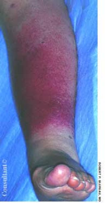
A 71-year-old man, who had recently returned from a month in Europe, complained of left lower leg swelling and pain of 1-week's duration. For many years, this obese patient had chronic venous insufficiency of both legs and chronic osteoarthritis of the knees that severely limited his ability to walk. The patient was admitted to the hospital with extensive cellulitis of the left lower leg.
An 11-year-old boy had generalized abdominal pain followed by nausea and vomiting. He had neither diarrhea nor fever. Examination revealed a flat, diffusely tender abdomen with no palpable masses. Pigmented macules were seen on his lips and buccal mucosa. A partial bowel resection had been performed 5 years ago because of intussusception.

Two days of pain in his right leg, which had been swollen for a week, brought this 69-year-old man with type II diabetes to the emergency department (ED). Three months earlier, the patient had undergone a radical retropubic prostatectomy with bilateral pelvic lymph node dissection. Examination in the ED revealed an edematous right leg indurated with a leathery-appearing thigh that was hot to the touch. His temperature was 38.7°C (101.7°F), and his white blood cell count was 21,290/µL with a shift to the left.
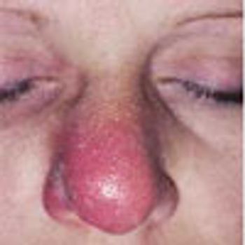
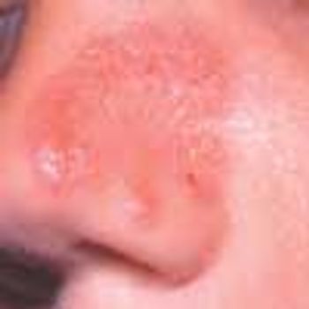
The nasal cellulitis that affects this 39-year-old woman began as right intranasal folliculitis. Because the patient was sensitive to many antibiotics, oral ciprofloxacin was prescribed.
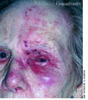
Periorbital, forehead, and nasal erythema, crusting, and pain typical of herpes zoster affected a 90-year-old woman. Reactivation of the latent varicella zoster is more common in the elderly and is attributed to impaired immunologic mechanisms.
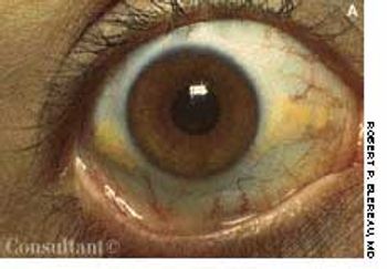
Pingueculae-yellow lipid deposits-may develop on the bulbar conjunctiva after several years of exposure to drying, dust, and the UV rays of sunlight. They represent elastotic degeneration of the substantia propria and are not seen in infants or children. These lesions usually are evident on the nasal aspect of the conjunctiva; they are innocuous and require no treatment.
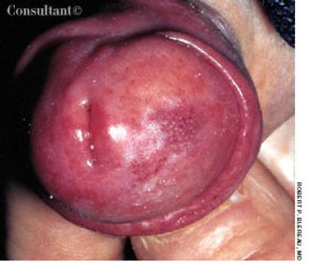
A 78-year-old man presented with an asymptomatic, irregular lesion on the right side of the glans penis. The 0.5-cm, light purple injected, finely papular lesion had spontaneously erupted about 2 months earlier.
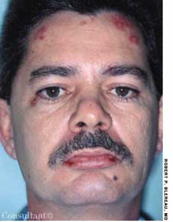
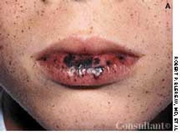
Numerous plaques, some with yellow crusting and central scarring, had erupted primarily on the face and neck of a 46-year-old man. A single lesion had developed on his left elbow as well. The lesions were initially diagnosed as impetigo, but they failed to resolve after 2 courses of oral cephalexin.
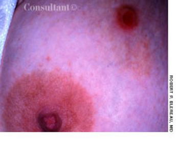
For several weeks, a 30-year-old woman had a 1 × 2-cm, oval lesion with a rolled border in the inner upper quadrant of the right breast that had progressed in size and ulcerated. Six weeks earlier, she had been bitten by her infant son in the same area. The medical history and physical examination were otherwise normal.
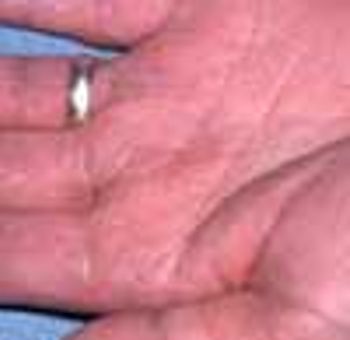
For about 4 months, a very dry, diffuse, fine scaly, asymptomatic eruption covered the palms of a 28-year-old man; several fingernails were dystrophic bilaterally as well. Before the onset of this condition, bilateral onychomycosis of the toenails had been diagnosed.

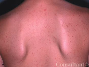
A 22-year-old man's chest shows the most common form of congenital pectoralis major muscle anomaly-partial absence of the right pectoralis major muscle with the clavicular origin intact.
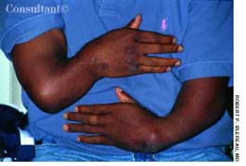
A 29-year-old man was born with radial clubhands that required several operations during his early weeks of life. In addition to shortened forearms, the patient was left with severe limitation of wrist motion bilaterally.
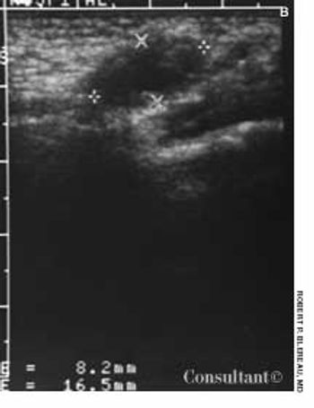
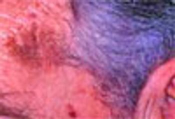
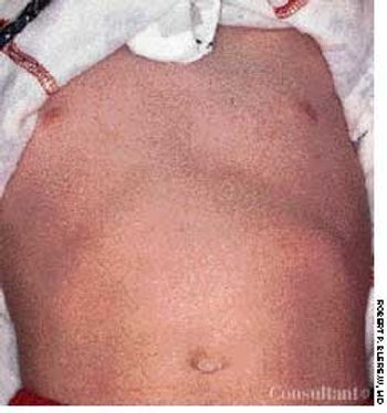
This 2-month-old infant was brought to her physician with a 2-week history of intermittent swelling in the right distal inguinal and upper labial area. A palpable solid, nonreducible lump was visualized ultrasonographically as an oval, hypoechoic mass and identified as a herniated ovary. The hernia was repaired surgically, and the ovary remained intact.