
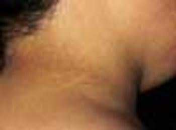
Hyperpigmentation involving the neck developed in this 8-year-old obese girl. The affected area resembled unwashed skin. The patient had worn a uniform to school-a jumper and blouse with a collar-for 6 months.
Hyperpigmentation involving the neck developed in this 8-year-old obese girl. The affected area resembled unwashed skin. The patient had worn a uniform to school-a jumper and blouse with a collar-for 6 months.
A mother, fearing that her 4-year-old son had been abused at his day-care center, rushed him to the emergency department, where an evaluation revealed a platelet count of 1,000/µL. Except for bruises on the boy's face and legs, the physical findings were normal. Bone marrow aspiration showed numerous megakaryocytes and was otherwise normal. The youngster's history included treatment for bronchitis, sinusitis, and conjunctivitis 2 weeks earlier.
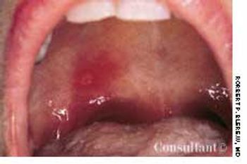
An afebrile 23-year-old man with no cervical gland enlargement presented with the very painful, large, shallow, yellowish-based ulcer shown here.

Erythematous, scaly lesions with double-edged borders had been present on a 14-year-old boy's left upper arm and lower legs for about a year. The lesions were occasionally pruritic, and some resembled ringworm. At times, fine yellow crusting suggestive of impetigo was present. The boy took very hot baths and showers.
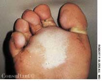
For 2 years, a 40-year-old oil-field worker wore heavy, steel-toed work boots all day. He experienced weeping and white discoloration at the center of both distal soles. At night, with his boots off, the feet would dry and hurt. There was no pruritus.
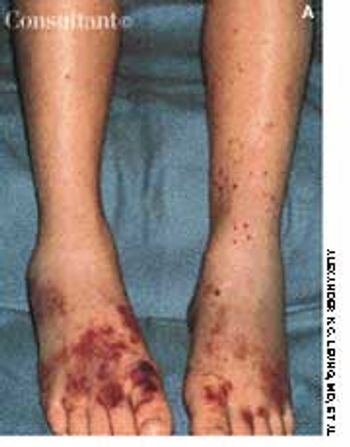
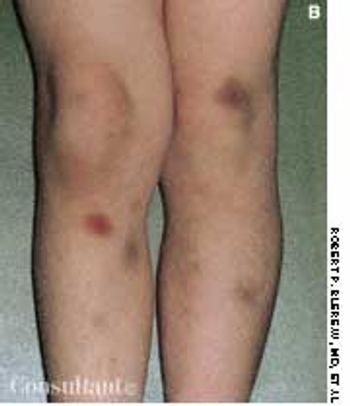
Two 7-year-olds show the purpuric rash of the lower body and legs that is typical of Henoch-Schönlein purpura. This disease is a vasculitis that chiefly affects small vessels of the skin, joints, gastrointestinal tract, and kidney.
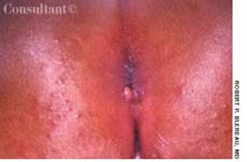
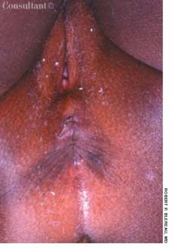
A 3-mm long, double-tipped, polypoid lesion appeared just anterior to the anus on an 8-month-old girl 2 days earlier. The lesion was excised in the office: a local anesthetic was administered, and the base was lightly electrodesiccated. Antibiotic ointment was applied until the area healed. Pathologic findings identified an infantile perianal pyramidal protrusion.

During a routine preschool examination, it was noted that this 4-year-old boy, who weighed 30.8 kg (68 lb), did not seem to rotate his arms. The child's mother stated that she first noticed this 2 or 3 years earlier but thought it was related to her son's obesity. The youngster appears to function normally; he is able to write and play football and basketball. No other family members are known to be similarly affected.
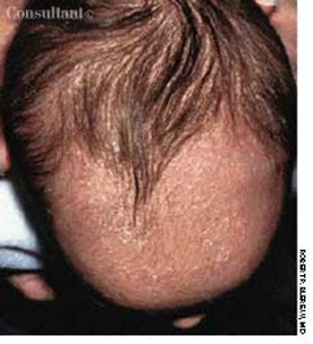
The entire scalp of this 1-month-old boy shows the diffuse yellow scaling of seborrheic dermatitis. Called “cradle cap” in infants, this dermatitis typically begins on the scalp during the first 3 months and may spread over the entire face. An associated stubborn diaper rash may develop in some infants.
A 56-year-old woman was bothered by “something protruding” from her vagina. She had no pain, bleeding, or dyspareunia.
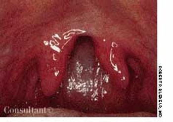
This striking picture of a bifid uvula in an asymptomatic 57-year-old man was taken by Robert P. Blereau, MD of Morgan City, La.
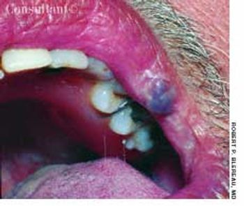
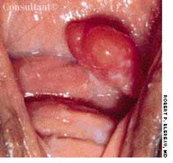
For 3 years, a gradually enlarging, raised, purple, cystic lesion had been present on the left upper lip vermilion of a 51-year-old man. The asymptomatic lesion measured 0.5 cm in diameter. The patient was given a local anesthetic, and the lesion was excised by wedge resection in the office; pathologic examination confirmed the diagnosis of benign cavernous hemangioma.
This asymptomatic lesion on the upper arm of a 60-year-old man had been present for 2 years. The patient had used several over-the-counter antifungal and hydrocortisone creams to treat what he thought was ringworm.
Enterobiasis-an infection by pinworms-is caused by the nematode Enterobius vermicularis. Treatment is a single dose of oral albendazole, mebendazole, or pyrantel pamoate.
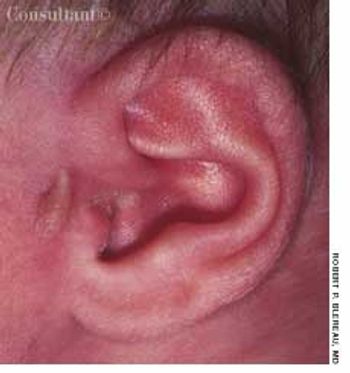
The left ear of a 1-month-old infant girl shows a congenital papillomatous lesion at the preauricular area. She had no other apparent abnormalities.
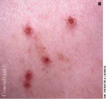
Highly pruritic, 2- to 4-mm, papular lesions with central ulceration erupted on the back of a 66-year-old woman. She had had 2 similar outbreaks in the past. The patient was taking conjugated estrogens, alprazolam, and alendronate.
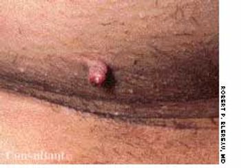
A 43-year-old woman requested removal of a lesion from her left axilla because it had recently become irritated. The polypoid lesion with dried terminal ulceration had been present for 17 years.
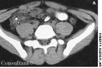
A 15-year-old boy presented with sharp, right lower quadrant abdominal pain, nausea, vomiting, and fever of 2 days' duration. His white blood cell count was 15,000/mL with a shift to the left on the differential. Right lower quadrant rebound tenderness was noted.
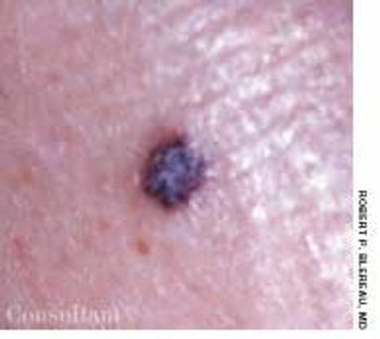
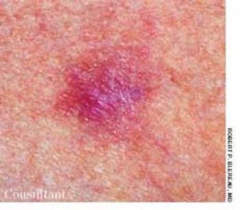
A 65-year-old man had had an asymptomatic lesion on his left lateral lower leg for several weeks. The dark maroon, almost black, 3- to 4-mm, circular, elevated lesion had a convoluted surface of dilated vessels.
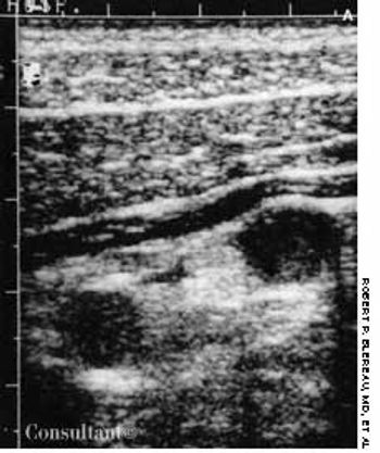
The diagnosis of acute appendicitis is usually straightforward. When the presenting symptoms are atypical, abdominal ultrasonography can be of diagnostic assistance if it shows the thickened walls of the appendix and a distended, noncompressible lumen.
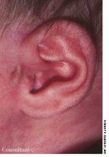
The left ear of a 1-month-old infant girl shows a congenital papillomatous lesion at the preauricular area. She had no other apparent abnormalities.
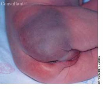
This photo was taken minutes after the birth of a female newborn and reveals a purple discoloration of the right buttock.
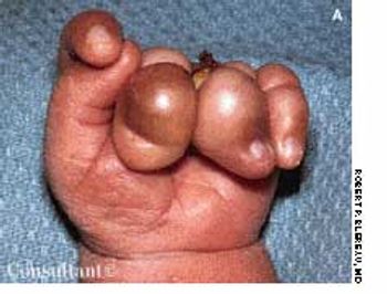
These photos are of the left hand of a full-term, newborn infant with focal ring constrictions of the third, fourth, and fifth fingers; secondary edema and discoloration are also seen.
Two days after beginning therapy with amlodipine, this 30-year-old woman noticed a pruritic, erythematous, macular dermatitis on her ankles.
These painful eczematous lesionsat the angle of the mouth and thebase of the nostrils had been presentin a 52-year-old woman for 3days (A). Some of the vesicles hadulcerated and left a crust over theregion. The patient said she had hadsimilar attacks in the past. The diagnosisof recurrent herpes simplexvirus 1 (HSV-1) infection was made.The patient was treated with acyclovirfor 1 week, and all the lesionsdisappeared.
A massive amount of blood in the inferior medial quadrantof the conjunctiva and ecchymosis of the lowereyelid of the right eye prompted a 60-year-old woman toseek medical attention. She had first noted blood in theeye 5 days earlier. There was no history of trauma.
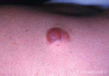
A 14-year-old girl was concerned about this 1-cm, red, nodulocystic lesion on her left posterior shoulder that had been present for several months. The lesion had developed over the site at which a 0.5-cm pilomatrixoma had been excised a year before. Four years earlier, another 1.9-cm pilomatrixoma had been excised from the girl’s right outer upper arm. There was no family history of the lesion.
A 36-year-old woman was concerned about the diffuseyellow pigment at the temporal aspect of the conjunctivaof her right eye.
For years, a 60-year-old obese woman with type 2 diabetes mellitus had thiswarty lesion on the outer tarsal plate in the middle of the left upper eyelid.It was asymptomatic. She also had a few asymptomatic epidermal inclusioncysts on the upper and lower eyelids.
The irides of a legally blind 19-year-old woman had been absent since birth.When she was 6 weeks of age, her parents noted that she was not focusingon objects the way her siblings had. They consulted an ophthalmologistwho diagnosed aniridia. The woman is able to read book print close up andcan ambulate independently, although she has difficulty at times, such aswhen stepping off a curb in unfamiliar surroundings.