
Brown pigmentation of the medial 60% of the left iris was noted in a 40-yearoldwoman. The remainder of the iris was blue, as was the entire right iris.She stated that she had a “spot” of brown in the left iris at birth. The pigmentedarea had gradually enlarged until puberty and had not changedsince then. Her vision was normal.
Robert P. Blereau, MD
Advertisement
Articles by Robert P. Blereau, MD
When palatal petechiae are present along with exudative tonsillitis and cervical adenitis, and test and culture are positive, a Strep diagnosis is more secure.
Hyperpigmentation is seen on the cheeks and eyelids of a 36-year-old woman.She became hyperthyroid at age 19 years, with accompanying exophthalmosand hyperpigmentation, following the birth of her first child. Thyroidectomywas carried out at that time, and the patient has been receiving thyroid replacementtherapy ever since. The hyperpigmentation, an uncommon accompanimentof hyperthyroidism, has persisted.
For more than 20 years, a 55-year-old man had a faintly erythematous, papulosquamousrash with arciform borders on his groin and waistline. The rashhad been treated with a variety of medications. Topical and oral antifungalsand antibiotics and topical corticosteroids had been used but to no avail. Nolaboratory tests had been performed.
For 2 months, an asymptomatic rash had been present on the upper arms of a 16-year-old boy of normal weight. The rash, as seen on the patient’s right arm, consisted of abundant fine papules. He had no other lesions.
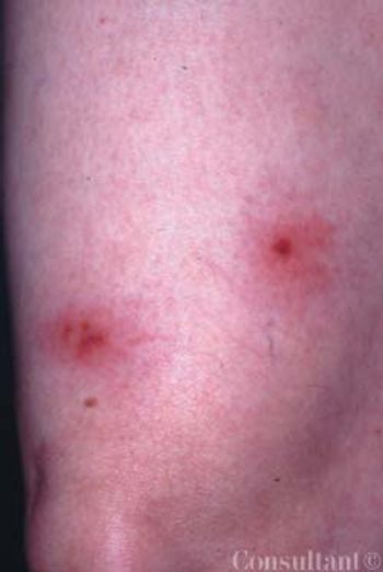
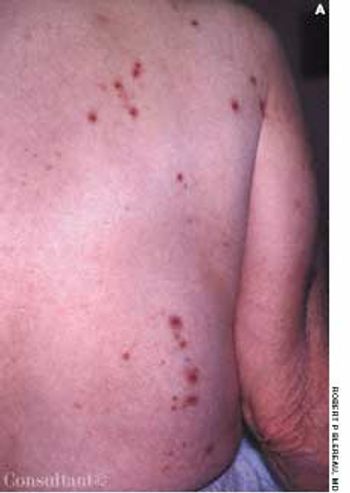
A 16-year-old girl was bothered byankle pain and “red spots” on herlower legs. These symptoms clearedin a few days without treatment. Sixweeks later, after returning from anall-day outing at a fair, she noticedthat the spots had reappeared (A)and hemorrhagic lesions had developedon the right ankle (B) and leftheel (C). After removing her shoes,the teenager felt severe pain in bothankles, particularly the right.
Sudden pain and a pulsatile swellingof the right upper medial thigh concernedan 80-year-old woman (A).Coronary angioplasty had been performedthrough this site 5 weeksearlier.
Large, reddish tan, asymptomatic patches recently developedon the trunk of a 36-year-old man. Five years earlier,a similar outbreak had resolved after a 10-day course oforal ketoconazole; this episode was the first recurrence.
A new lesion recently arose on the right flexor forearm ofa 67-year-old man. The 1-cm, pruritic, pink, circular, slightlyraised lesion was perfectly homogeneous with no centralclearing.
A 49-year-old man was concerned about a right flexor forearmlesion that had been increasing in size for 6 weeks.The light pink, well-demarcated, 5-cm, circular lesion featuredslight peripheral elevation with ulceration, crusting,and a relatively clear central area. A culture of materialfrom the lesion was negative for fungi. A potassium hydroxideevaluation was not performed.
A 56-year-old man had an asymmetric,maculopapular, sharply demarcated,pruritic, excoriated dermatitis on hisupper thighs. The eruption had beenpresent for 2 to 3 weeks
For about 4 months, a very dry, diffuse,fine scaly, asymptomatic eruptioncovered the palms of a 28-yearoldman; several fingernails weredystrophic bilaterally as well. Beforethe onset of this condition, bilateralonychomycosis of the toenails hadbeen diagnosed. The toenails had notbeen treated and were still affectedat the time of presentation. Branchinghyphae were seen on a potassiumhydroxide preparation of a fingernailcutting. The patient had tinea manuumand tinea unguium
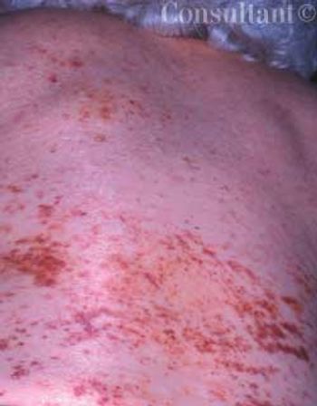
Diffuse petechiae suddenly arose on the back and abdomen of a 79-year-old woman. Within several days, the asymptomatic lesions covered her arms and face as well.
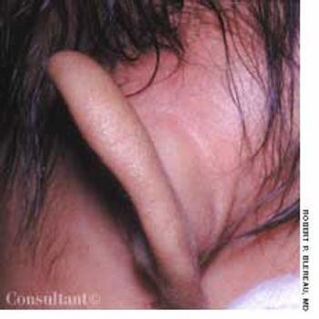
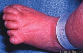
Concerned that her 7-week-old daughter's left ear was far more prominent than the right one, the mother took the infant to the emergency department (ED) for evaluation. The swelling had begun 3 or 4 days earlier; the patient was otherwise asymptomatic.
The 2-mm, white, very slightly raised lesion on the vermilion of a 37-year-oldman’s lower lip had been present for 2 months. The lesion was asymptomatic.There was no history of injury or burn to the area.
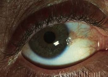
Pingueculae represent elastotic degeneration of the substantia propria.They develop on the bulbar conjunctiva of older persons after prolonged exposure to drying, dust, and the UV rays of the sun.
This asymptomatic lesion had been present on a 75-year-old man's right buttock for 2 to 3 years. The 0.5 cm in diameter nodule featured an irregular, flesh-colored surface.
A 1-cm, tan-brown lesion had developed years earlier at the posterior helix of a61-year-old man’s right ear. Central ulceration and crusting were noted on thepapule. The patient sought medical evaluation when the lesion became nodularand began to flake.
A 65-year-old woman presents with a 1-cm raised, light brown, circular, nodularlesion on the top of her head, which has progressively enlarged during the last6 months. The growth was removed by shave excision; electrodesiccation andcurettage were performed on the base. Pathologic evaluation determined thelesion was a basal cell carcinoma.
This 2-day-old infant was noted by Robert P. Blereau, MD, of Morgan City, La, to have duplication of the fifth toe with webbing between the two toes to the distal interphalangeal joint. There was a family history of polydactyly on the paternal side and syndactyly on the maternal side.
Several lesions had appeared 1 week earlier on the left distal thigh of a 47-year-old woman. The affected area featured erythema with irregular faded borders and central redness with very fine papules over the L3 dermatome. The indurated and tender central areas suggested inflammation or necrosis. There were no vesicles and there was no drainage.
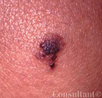
This lesion on her knee had been present for 5 years, reported a 22- year-old woman. It was not related to any trauma; its size had not changed, but occasionally it became darker or lighter.

Round, smooth, bald areas had recently developed on the neck and chin of a 32-year-old man. No other areas of the body were affected. The patient had a lifelong history of chronic anxiety.
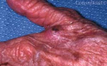
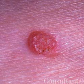
An 87-year-old woman sought treatment of what she described as a “bite” of 1 month’s duration. The pink, nodular lesion on the dorsum of the left hand had central superficial ulceration with yellow crusting at the web space between the thumb and index finger.
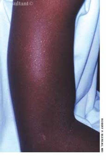
Some degree of hyperpigmentationdevelops in most pregnantwomen. This coloration is more pronouncedin dark-skinned women; onnaturally pigmented areas, such as theareolae, perineum, and umbilicus; andon the axillae, inner thighs, and otherregions that are prone to friction.
Approximately 2 weeks earlier, a pruritic,papular eruption had developed overthe abdomen of a 33-year-old womanwho was 34 weeks' pregnant. Therash was confined mainly to the striaedistensae (Figure 3). Because thesite was severely pruritic, the patientwas unable to sleep. Based on theclinical presentation, pruritic urticarialpapules and plaques of pregnancy(PUPPP) was diagnosed.
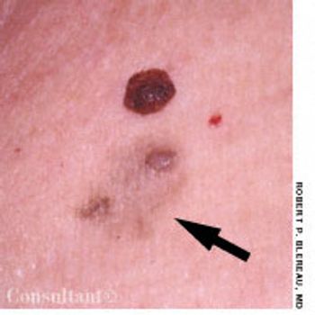
Two asymptomatic lesions that had been present for 1 year on the left upper chest of a 61-year-old man were excised. Pathologic examination revealed the raised, dark brown medial lesion to be an interdermal nevus; the lateral, light tan lesion with papules was a papillomatous interdermal nevus with primary macular amyloidosis.
Highly pruritic, 2- to 4-mm, papular lesions with central ulceration erupted on the back of a 66-year-old woman. She had had 2 similar outbreaks in the past. The patient was taking conjugated estrogens, alprazolam, and alendronate.

Numerous plaques, some with yellow crusting and central scarring, had erupted primarily on the face and neck of a 46-year-old man. A single lesion had developed on his left elbow as well. The lesions were initially diagnosed as impetigo, but they failed to resolve after 2 courses of oral cephalexin.
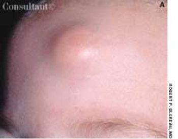
A 0.5-cm mass with overlying erythema on the forehead of a 5-monthold girl was first noticed after she had been hit on the head with a rattle. Excision of the mass was deferred because it was considered to be a hemangioma.
Advertisement
Advertisement
Advertisement
Trending on Patient Care Online
1
First Oral Film Treatment for Erectile Dysfunction in Men Gains FDA Approval
2
Long-Term Data Support Sustained Bimekizumab Response in Hidradenitis Suppurativa
3
Weekly Dose Podcast: New Obesity Data, Insulin Guidance, and Mental Health Screening
4
Topline Phase 2 Data Show Roflumilast Cream Improves Atopic Dermatitis in Infants as Young as 3 Months
5