


Some sexually transmitteddiseases (STDs), such assyphilis and gonorrhea, arecenturies-old scourges; othershave attained clinicalsignificance only in recent years.Despite the availability of effectivetherapy for many of these diseases,they remain an important publichealth problem.
Dermatology
Latest News
Advertisement
Advertisement
Abstract: Failure to recognize heparin-induced thrombocytopenia (HIT) can lead to devastating thrombotic events, including pulmonary embolism and stroke. In most cases, the problem develops within 5 to 14 days after a first-time exposure to heparin. HIT can occur with either unfractionated heparin or low molecular weight heparin (LMWH), but the incidence is much lower with LMWH. When HIT is suspected clinically, a functional assay and immunoassay should be performed. However, treatment should not be delayed while waiting for laboratory confirmation. All forms of heparin should be eliminated, and treatment with an alternative anticoagulant should be initiated to prevent new thromboembolic events. Argatroban and lepirudin are the direct thrombin inhibitors that have been approved for the treatment of HIT. Because of the risk of warfarin-induced venous limb gangrene or skin necrosis, warfarin should be avoided in patients with acute HIT until their platelet counts have recovered and they are improving clinically. (J Respir Dis. 2006;27(6):248-259)
Standard first aid for insect bitesand stings involves cleansing the skinwith soap and water followed byapplication of an antiseptic and topicaldiphenhydramine.
Green discoloration of the fingernailsdeveloped 6 weeks after a 29-year-oldwoman had artificial nails placed duringa manicure. The patient was a doctorof pharmacy degree candidate whowas married and had 2 children.
A 24-year-old woman complained of a rash on both feet and legs. She had also had intermittent pain in both ankles for the past year.
For the past few weeks, pruriticpatches have been erupting on a38-year-old man’s extremities. He recallsthat similar lesions occurredduring the last 2 winters. The patienthas a history of seasonal allergies;he owns a cat and 2 dogs.
A 6-year-old girl presents witha several-month history ofgenital discomfort that includesitching, irritation, andoccasional bleeding. Themother reports that there isblood on the toilet paper afterthe child wipes herself. Therehas also been some spottingin the child’s underwear. Thepatient seems to be grabbingat her crotch frequently.
To reduce the incidence of candidalinfections in patients with dermatologicproblems, instruct them to drythe affected area with a blow-dryeron a cool or warm setting before theyapply topical medications.
Test your knowledge of dermatology with 4 cases, 4 photos, 4 different rashes . . . which are the result of a drug reaction?
In 2 recent “Dermclinic” cases (CONSULTANT, December 2001, page 1812),Dr David Kaplan describes young women with lesions that arose on their extremitiesafter they used hormonal agents:
A 32-year-old woman tells you that she has had generalized weakness;swelling of the face, arm, and legs; diffuse myalgias; and a facial rashfor several weeks. Her main concern is her inability to keep her arms elevatedor get out of bed. Her history is significant for cervical cancer.
A 79-year-old woman with a 37-year history of type 2 diabetes mellitus complains of head pain that began more thana month ago and is localized to the left frontotemporal region. She characterizes the pain as constant and burning, with minimalfluctuations in intensity. The pain does not increase with any particular activity but is quite disabling; it has causedemotional lability and insomnia. She denies nausea, visual disturbances, weakness of the extremities, dizziness, or tinnitus.Her appetite is depressed; she has experienced some weight loss.
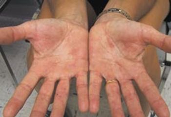
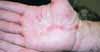
For 8 months, a 50-year-old woman had had an erythematous, pruritic rash on her palms. When her hands were exposed to water, the rash worsened and fissures developed. Recently, she noticed that the fissures had begun to bleed. Application of clobetasol, 0.05%, for 1 month provided no relief.
An 89-year-old hemiplegic man is seen for a routine physical examination. Major right brain stroke produced permanent dense left-sided motor deficit years ago. Has related vascular dementia.
A persistent, 2-month-old rash under both breasts has not responded to overthe-counter antifungal creams. The 55-year-old patient now seeks medical care;she is otherwise healthy.
My patient, a 33-year-old man who works with his hands, presented with a growthunder his left thumb nail (Figure).
We read with great interest the “Dermclinic” case of a 44-year-old man with achanging pigmented lesion on his trunk (CONSULTANT, March 2001, page 354).
The sudden onset of asymptomatic red streaks on several sites alarms a14-year-old girl. The patient is otherwise healthy; she denies any symptoms ofdepression.
A 62-year-old woman was admitted to the hospital with back pain, fatigue, andan ulcerated lesion on the anterior left foot. Clinical and laboratory findingsconfirmed the diagnosis of multiple myeloma.
An ulcer was noted on the right arm ofa 65-year-old woman with ulcerativecolitis who was being evaluated for apartial bowel resection. Antibiotic therapygiven 2 months earlier had no impacton the lesion; subsequent debridementonly increased the ulcer’s size.
This painful, eroded plaque on thedorsum of a 39-year-old man’s handhad developed over a few days from asmall, painful pustule. The patient’shistory included ulcerative colitis,which was not active when the lesionoccurred.
The pyoderma gangrenosum on theright anterior tibial area of a 40-yearoldman was thought to be associatedwith his rheumatoid arthritis. However,the cause of many of these ulcersis unknown. The patient could not recallany recent trauma. At least half ofall pyoderma gangrenosum lesionsoccur in persons who do not have associateddiseases.1
A tiny papule that arose after minortrauma to her finger marked theonset of this lesion, according to the48-year-old patient. She reports thatthe papule rapidly evolved into apustule that grew within 2 weeks intoa painful, undermined, purple-edgedulcer. The lesion did not respond toantibiotic therapy. The patient had rheumatoid arthritis.
Pyoderma gangrenosum is frequentlyassociated with systemic diseases,such as ulcerative colitis and Crohn’sdisease (Table). The occurrence of theskin ulcers does not necessarily correlatewith the activity of the underlyingdisorder.
A 57-year-old man was referred forevaluation of an enlarging, painful,irregular ulceration on his lower abdominalwall. The patient recalledhaving a small, red, “blister-like” lesionthat had rapidly expanded to itscurrent size of 2.5 * 4.5 cm. Hedenied specific injury to the skin;however, he often wore jeans thatrubbed the area. The patient wastaking ibuprofen for seropositiverheumatoid arthritis.
Advertisement
Advertisement
Trending on Patient Care Online
1
From Amyloid Clearance to Daytime Function: Why Sleep Quality Matters for Brain Health
2
2026 Dyslipidemia Guideline Expands Statin Eligibility to 21.5 Million More US Adults
3
ACOG Releases New Guidance on HIV Screening and Prevention
4
Compulsive Smartphone Use Linked to Depressive Symptoms in Older Adults
5