Cirrhosis and ascites developed in a 52-year-old man with a history of chronic hepatitis C and ethanol abuse. He was hospitalized because of bleeding esophageal varices, which were successfully treated with elastic band ligation.
All News
Advertisement
Advertisement
Lichen planus is characterized by flat-topped, polygonal, purple pruritic papules that have a predilection for flexor aspects of the wrists and forearms, sides of the neck, thighs, shins, and lower back. Lesions on the oral mucosa appear as white, lacy patches.
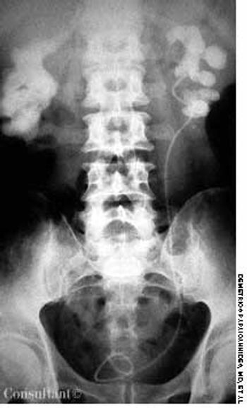
A roentgenogram of the kidneys, ureter, and bladder of a 58-year-old man shows bilateral stones in the renal pelvis and the renal calyces. The patient had a history of recurrent urinary tract infections caused by Proteus mirabilis. A ureteral catheter (pigtail) had been placed in the pelvis of the left kidney to facilitate drainage.

Skull x-ray films were taken after this 62-year-old nursing home resident fell and hit his head. The radiographs revealed only a sharply demarcated radiolucent area, mainly over the right parietal bone. Osteoporosis circumscripta was diagnosed.

A week after the onset of headache, fever, chills, nausea, weakness, and malaise, a 23-year-old man presented to the emergency department of a hospital on Long Island in New York. He reported that analgesics had not eased his symptoms. The patient's only past hospitalization was a splenectomy secondary to an auto accident at age 16.

A 40-year-old woman presented for follow-up of a generalized skin condition that was most severe on her palms and soles. She had been born with a few lesions, which resolved in infancy. New lesions began to emerge and increase in number and severity when she was 2 years old; they have recurred intermittently for 38 years.
Gonococcal infection is the leading cause of bacterial arthritis in adults.

The extent of mucosal or transmural intestinal necrosis varies. Pneumatosis progresses from the submucosa through the muscular layer to the subserosa. The distal ileum and proximal colon are most frequently involved.

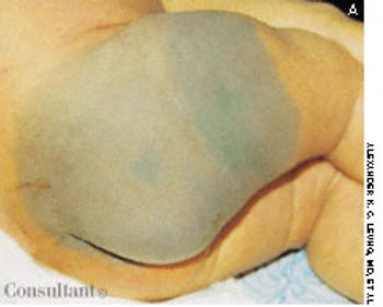
Mongolian spots are congenital, hyperpigmented, usually gray areas of varying size and shape. They result from the abnormal occurrence of melanocytes in the lower half of the dermis and are found most frequently in the sacrococcygeal and gluteal areas.
A 56-year-old man complained of perianal swelling and discomfort. He reported a history of diarrhea and mucus discharge from the rectum with intermittent rectal bleeding. A colonoscopy confirmed the diagnosis of Crohn's disease of the rectum and sigmoid colon.
Advertisement

A 30-year-old man presented to the emergency department with new-onset seizures. His past medical history included loss of vision for 1 year, deafness, and osteomyelitis of the mandible.

The initial complaint of a 79-year-old woman was of mild headache, neck pain, and sore throat. She had a history of hypertension, diabetes mellitus, and heavy cigarette smoking. Examination by an otolaryngologist, which included laryngoscopy, revealed no abnormalities. Three weeks later, the patient's throat and neck pain became more severe. She had no arthralgias, visual loss, fever, or worsening head pain.

Inspissated, sticky, immobile meconium causes this transient form of distal colonic or rectal obstruction in newborns. The incidence has been estimated at 1 in 500 to 1000 live births. The condition is thought to result from dehydration of the meconium.
These are hyperpigmented, regularly bordered, sharply demarcated macules that are usually tan or light brown in whites and dark brown in dark-skinned persons. The lesions are characterized by an increased number of melanocytes and an increased amount of melanin in the epidermis.
A 43-year-old woman was hospitalized with a 3-day history of fever and back pain. She was malnourished and seropositive for HIV infection. Results of blood and sputum cultures were negative. A community-acquired pneumonia was diagnosed. Chest film findings and the clinical presentation were inconsistent with Pneumocystis carinii pneumonia.
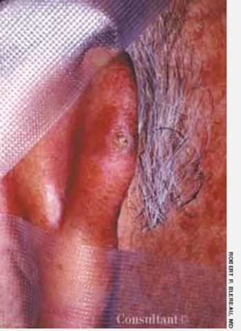
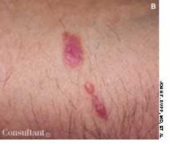
A 1-cm, tan-brown lesion had developed years earlier at the posterior helix of a 61-year-old man's right ear. Central ulceration and crusting were noted on the papule. The patient sought medical evaluation when the lesion became nodular and began to flake.
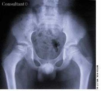
A 10-year-old boy presented with right hip pain and a limp. The patient was taking no medications and had no personal or family history of disease. He denied a history of trauma.
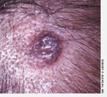
A 65-year-old woman presents with a 1-cm raised, light brown, circular, nodular lesion on the top of her head, which has progressively enlarged during the last 6 months.
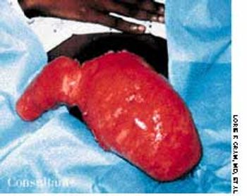
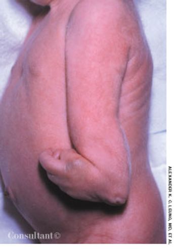
A firm mass projected from the deltoid muscle region of a 24-year-old man's right shoulder.
This newborn has trisomy 18, as manifested by intrauterine growth retardation (birth weight, 2350 g; length, 47.2 cm), microcephaly (head circumference, 31.5 cm), short neck, hypotonia, feeding difficulties, high-pitched cry, micrognathia, cleft palate, low-set ears, short sternum, widely spaced nipples, clenched hands with ulnar deviation and overlapping digits, micromelia, and a single umbilical artery.
Advertisement
Advertisement
Advertisement