All News

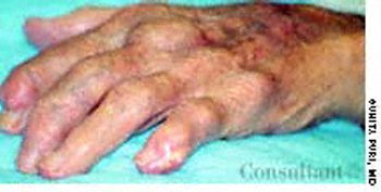
A 65-year-old man had a 10-year history of deformity of the hands, pain, and nodules on the fingers. His serum uric acid level was 9 mg/dL. The suspected diagnosis of chronic tophaceous gout was confirmed by the finding of birefringent monosodium urate needle-shaped crystals in the joint fluid and the patient's significant response to colchicine within 12 to 24 hours of the start of therapy.
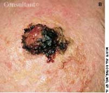
An 82-year-old man with Alzheimer's disease and atrial fibrillation was referred for evaluation of a lesion present for an undetermined period. It was initially noted 1 week earlier, at his first office visit with a geriatrician.

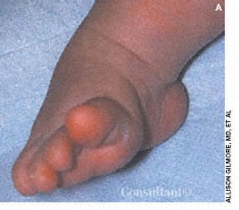
Congenital Clubfoot

This 3-month-old boy has an untreated right clubfoot deformity. The foot is stiff and uncorrectable. The vertical midfoot crease indicates significant deformity. An anteroposterior (AP) simulated weight-bearing radiograph shows parallelism of the long axis of the talus and calcaneus; this indicates varus alignment of the hindfoot. There is also forefoot adduction.

Bilateral Cryptorchidism

The mother of a 2-year-old boy was concerned because his scrotum appeared flat and empty. (She had noticed this earlier but thought it was normal for her child's age.) On examination, the testicles were not palpable in the scrotum. They were found in the inguinal area and could not be manipulated into the scrotum. Bilateral orchidopexy was performed, and the postoperative course was uneventful.

Leukocytoclastic Vasculitis

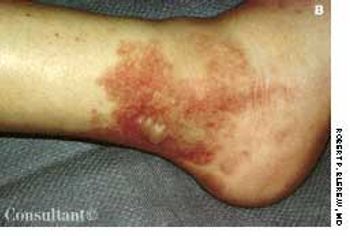
A 16-year-old girl was bothered by ankle pain and “red spots” on her lower legs. These symptoms cleared in a few days without treatment. Six weeks later, after returning from an all-day outing at a fair, she noticed that the spots reappeared and hemorrhagic lesions developed on the right ankle and left heel. After removing her shoes, the teen-ager felt severe pain in both ankles, particularly the right.

Livedo reticularis is the cutaneous manifestation most frequently associated with the antiphospholipid syndrome (APS). It manifests as a dusky, violaceous vascular discoloration with a reticulated pattern on the upper and lower extremities.

Dislocated Septal Cartilage

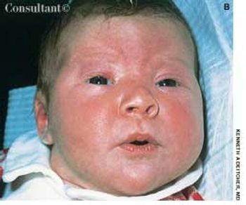
Dislocation of the triangular cartilage of the nasal septum is caused by pressure on the nose before and during delivery. It should not be confused with the transient flattening of the nose during birth.

Colonic Adenoma

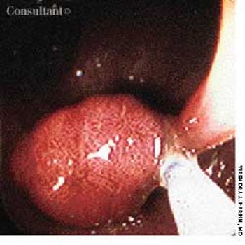
When a 59-year-old man's stool tested positive for occult blood, colonoscopic evaluation was carried out. This is the endoscopic appearance of a pedunculated colonic adenoma-the source of the bleeding-being removed by a snare.

Snakeskin Gastric Mucosa

Cirrhosis and ascites developed in a 52-year-old man with a history of chronic hepatitis C and ethanol abuse. He was hospitalized because of bleeding esophageal varices, which were successfully treated with elastic band ligation.

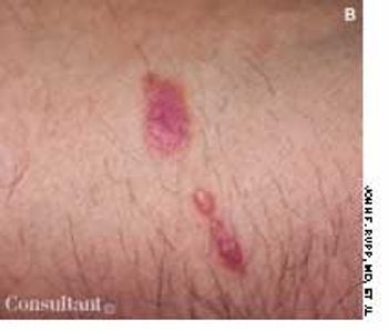
Lichen planus is characterized by flat-topped, polygonal, purple pruritic papules that have a predilection for flexor aspects of the wrists and forearms, sides of the neck, thighs, shins, and lower back. Lesions on the oral mucosa appear as white, lacy patches.

Bilateral Staghorn Calculi

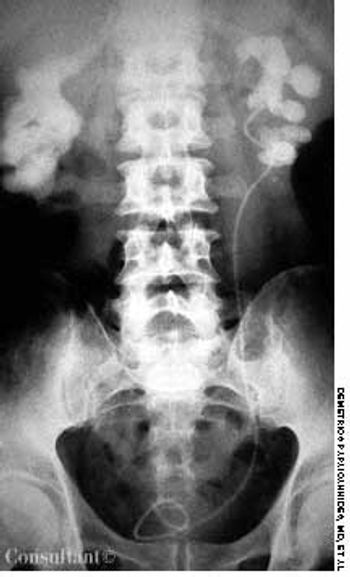
A roentgenogram of the kidneys, ureter, and bladder of a 58-year-old man shows bilateral stones in the renal pelvis and the renal calyces. The patient had a history of recurrent urinary tract infections caused by Proteus mirabilis. A ureteral catheter (pigtail) had been placed in the pelvis of the left kidney to facilitate drainage.

Osteoporosis Circumscripta

Skull x-ray films were taken after this 62-year-old nursing home resident fell and hit his head. The radiographs revealed only a sharply demarcated radiolucent area, mainly over the right parietal bone. Osteoporosis circumscripta was diagnosed.

Babesiosis

A week after the onset of headache, fever, chills, nausea, weakness, and malaise, a 23-year-old man presented to the emergency department of a hospital on Long Island in New York. He reported that analgesics had not eased his symptoms. The patient's only past hospitalization was a splenectomy secondary to an auto accident at age 16.

Epidermolysis Bullosa

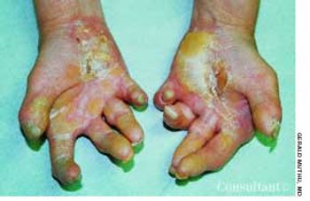
A 40-year-old woman presented for follow-up of a generalized skin condition that was most severe on her palms and soles. She had been born with a few lesions, which resolved in infancy. New lesions began to emerge and increase in number and severity when she was 2 years old; they have recurred intermittently for 38 years.

Bacterial Arthritides

Gonococcal infection is the leading cause of bacterial arthritis in adults.

Necrotizing Enterocolitis

The extent of mucosal or transmural intestinal necrosis varies. Pneumatosis progresses from the submucosa through the muscular layer to the subserosa. The distal ileum and proximal colon are most frequently involved.

Salmon Patches

Mongolian spots are congenital, hyperpigmented, usually gray areas of varying size and shape. They result from the abnormal occurrence of melanocytes in the lower half of the dermis and are found most frequently in the sacrococcygeal and gluteal areas.

Perianal Crohn's Disease

A 56-year-old man complained of perianal swelling and discomfort. He reported a history of diarrhea and mucus discharge from the rectum with intermittent rectal bleeding. A colonoscopy confirmed the diagnosis of Crohn's disease of the rectum and sigmoid colon.

Osteopetrosis

A 30-year-old man presented to the emergency department with new-onset seizures. His past medical history included loss of vision for 1 year, deafness, and osteomyelitis of the mandible.

Lingual Artery Infarct

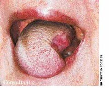
The initial complaint of a 79-year-old woman was of mild headache, neck pain, and sore throat. She had a history of hypertension, diabetes mellitus, and heavy cigarette smoking. Examination by an otolaryngologist, which included laryngoscopy, revealed no abnormalities. Three weeks later, the patient's throat and neck pain became more severe. She had no arthralgias, visual loss, fever, or worsening head pain.