
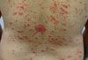





















































































































































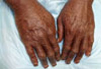
Prominent, pigmented polyangular scales on the extensor surfaces of the arms, legs, and trunk suggested recessive X-linked ichthyosis. DNA analysis demonstrated an abnormal steroid sulfatase gene, which confirmed the diagnosis.
Prominent, pigmented polyangular scales on the extensor surfaces of the arms, legs, and trunk suggested recessive X-linked ichthyosis. DNA analysis demonstrated an abnormal steroid sulfatase gene, which confirmed the diagnosis.
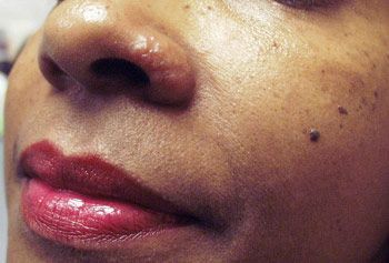
Test your clinical skills in this week’s 5-question quiz.
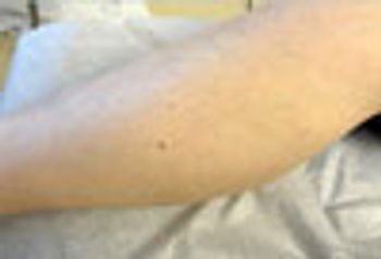
Eccentric darker pigmentation in this small asymptomatic lesion strongly suggests malignant melanoma. This patient had a history of non-melanoma skin cancer, but was unaware of the questionable growth.

This is a classic keloidal mass, which can occur in any ethic group. Because keloids can be intensely painful, they almost always require removal.
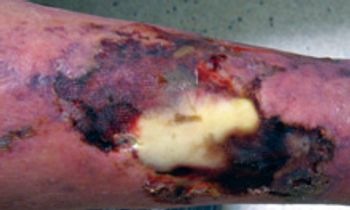
Preseptal cellulitis, myasthenia gravis, squamous cell carcinoma, atheromatous embolism, calciphylaxis are shown here.
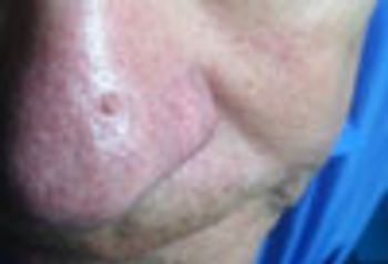
Histopathology showed this neoplasm to be a seborrheic keratosis: it might well have been a hypertrophic actinic keratosis, melanoma, or superficial basal or squamous cell carcinoma.
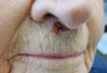
Seborrheic keratosis; retropharyngeal abscess; in situ malignant melanoma; statin-induced myalgias; basal cell carcinoma
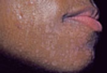
Facial papules; pediatric gastrostomy complications; unexplained trismus; renal abscess.
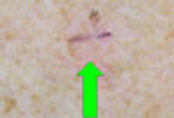
Test your clinical skills in this week’s 5-question quiz.
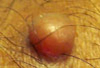
Carcinoma?; spirometry to assess chronic cough?; neurofibroma?; laryngeal neoplasm? What do you think?
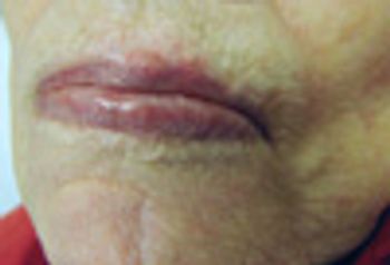
Diabetic myonecrosis; Persistent bacterial bronchitis; basal cell carcinoma; statin-drug pleiotropy; spinal epidural abscess
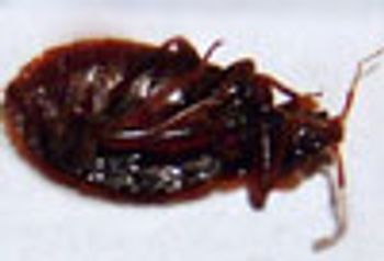
This patient wonders whether the insect he found on his headboard might be responsible for the clusters of pruritic, crusted papules on his legs. This turned out to be a bed bug.
The areas that contained linear erythema, punctuated by stippled pigmentation, and a small round area of stippled pigmentation were biopsied: both showed pigmented basal cell carcinoma.
This new-onset “growth” was removed and shown to be a solitary neurofibroma, which is not an indication of any of the genodermatoses marked by a multiplicity of such neoplasms.
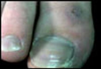
Acne?; Halo Nevus; Solitary Fibrous Tumor; idiopathic chilblains
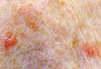
Biopsy of the 3 red “spots” in close proximity showed lichenoid keratosis-a benign lesion, probably related to seborrheic keratosis, which often mimics skin cancer.
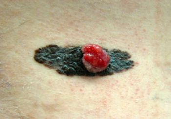
Livedo reticularis; persistent fatigue and weight gain; groin rash; paraneoplastic syndrome; funny mole.
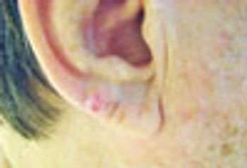
While it might be tempting to dismiss this as a picker's nodule, lesion's thickness suggested the need for a biopsy, which revealed that the lesion was an infiltrative, invasive basal cell carcinoma. The lesson? Never hesitate to biopsy when the slightest uncertainty exists as to the correct diagnosis.
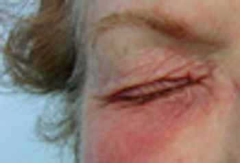
The predominantly infraorbital erythema and swelling suggested that this rash was related to a material that was being placed in the eyes and dripping from them. Patch testing showed that the patient was allergic to the preservative in the eyedrops she had been using.
The translucent papule on the upper lip has a few small blood vessels that traverse its surface. These are morphologic features of basal cell carcinoma, a presumptive diagnosis that was confirmed by biopsy.
Does this look like melanoma? Which is not true of Löfgren syndrome? Yoga and Afib; what cause of mental confusion? Skin lesions and HIV.. . . See if you can answer this week's quiz questions.
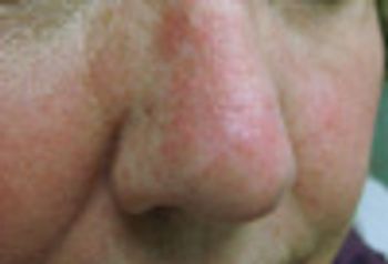
The redness on this woman's nose represents fairly severe photodamage with extensive actinic keratosis. Use of a topical therapy to achieve clearance of a field of actinic damage can be done with imiquimod, 5-fluorouracil, or ingenol mebutate creams, or diclofenac gel.
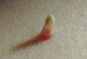
Cutaneous horns can arise on top of: seborrheic keratosis (as in this patient’s case), actinic keratoses, warts, basal and squamous cell carcinomas. Therefore, the lesion-and especially the base-must be submitted for pathologic diagnosis.
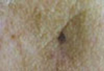
“Stippled” pigment is highly typical of a pigmented basal cell carcinoma. Diagnostic contenders include atypical seborrheic keratosis and superficial spreading melanoma.
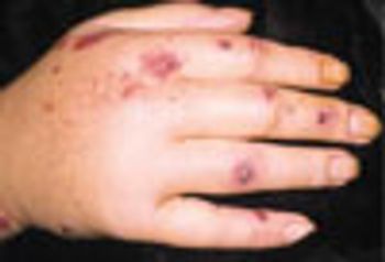
Edema of the hands has numerous etiologies: leukocytoclastic vasculitis, puffy hands from HCV infection, DVT, lymphedema, trauma