

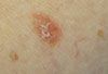


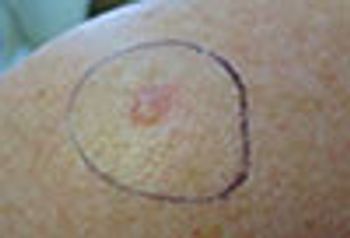
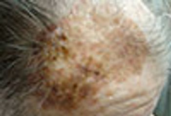
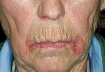
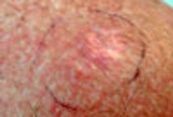
Biopsy of the flat area of this erythematous patch revealed superficial basal cell carcinoma while biopsy of the more nodular component revealed deeply infiltrating basal cell carcinoma.
Biopsy of the flat area of this erythematous patch revealed superficial basal cell carcinoma while biopsy of the more nodular component revealed deeply infiltrating basal cell carcinoma.
An asymptomatic bump; IBD and celiac disease; a gradually expanding spot .... test your clinical skills on a range of disorders in this week's quiz.
Add skin health to the list of medical conditions that are affected by poor sleep. Sun protection should be emphasized in skin care.
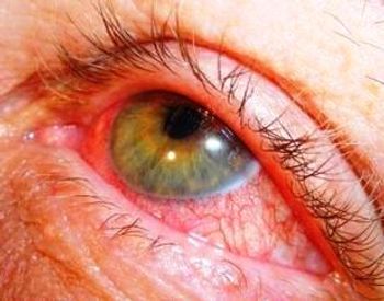
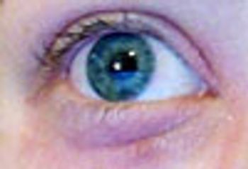
The eyes may be "windows to the soul," but they also provide a look at your patients’ overall health and clues to specific disorders. This week’s photo essay will test your ability to identify eye-related problems.
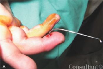
What's the best way to dislodge a fishhook? What if there is bony involvement?
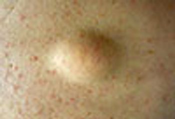
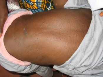
This non-tender, freely mobile, rubbery-feeling nodule is a classic lipoma. Diagnostic contenders include epidermal cyst and cutaneous metastases.
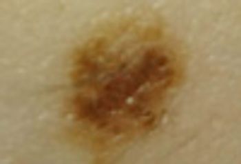
Eccentric and irregular pigmentation prompted a biopsy in this elderly woman with a history of basal cell carcinoma and melanoma. The surprising diagnosis was a heavily pigmented squamous cell carcinoma in situ.
Creatures of all kinds can get under your patients’ skin and cause problems, and the results sometimes are mistaken for other types of lesions. This week’s photo essay will test your ability to identify various kinds of diagnoses.
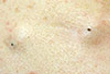
The puncta, with a black center, clearly identified these as classic epidermal cysts.
Ulcerating ankle; loud snoring; a new onset rash: see if you can answer this week’s 5 quiz questions.
Biopsies of this large variegated pigmented plaque ruled out lentigo maligna melanoma and gigantic seborrheic keratosis.
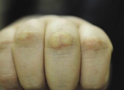
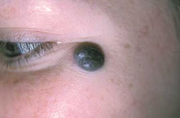
This translucent dark pink nodule located just behind the nail is the classic presentation of a digital myxoid cyst.
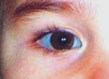
A rapidly developing lesion . . . flashing lights . . . blue sclera: can you answer this week’s 5 quiz questions?
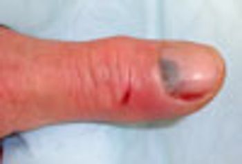
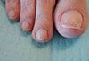
After a dog bite, trauma in the form of a subungual hematoma and shallow lacerations prompted a 10-day course of amoxicillin-clavulanate. This antibiotic is also useful for cat and human bites that appear to be at high risk of becoming infected.
Read carefully this week: you'll find helpful clues in at least 3 of the descriptions . . .
Here: short cases with photos that show melanoma, and pigmented lesions that mimic melanoma.
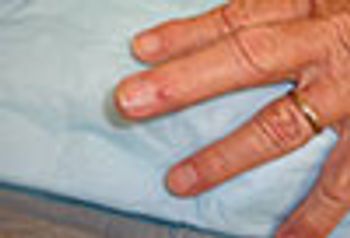
A biopsy revealed non-caseating granulomata, and culture revealed Mycobacterium marinum. This patient had a fish tank at home, and used his right hand to perform maintenance.
Go for the Glory Quiz - Does this rash suggest something more than skin deep? Diabetes and CKD? Scrotal lesions? See if you can answer all 5 quiz questions.
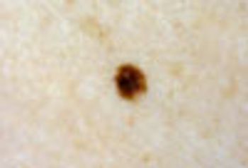
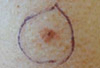
This pigmented macule clearly stands out against a bland background of fair-complected skin. This is known as the “ugly duckling” sign. Such lesions should always be viewed with suspicion and a biopsy is generally indicated.
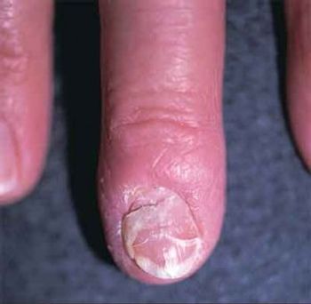
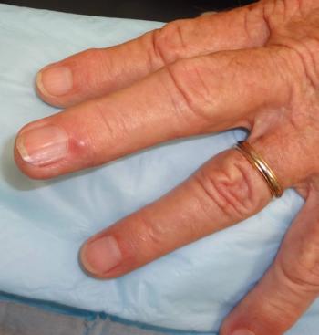
Is thyroid hormone medication associated with nail plate deformity, or is something else going on?
This tumor is a low-grade soft tissue sarcoma. Metastases are uncommon, but local recurrence is very frequent. Mohs surgery is the optimal treatment modality.
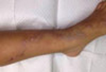
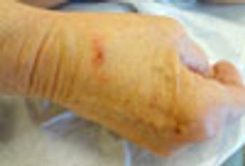
Cutaneous larva migrans (CLM), also known as “creeping eruption,” is the most commonly acquired tropical dermatosis
Recent trauma caused this lesion, which had been present since the patient’s birth, to enlarge. What is it?
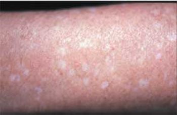
Images of guttate hypomelanosis, jellyfish sting, basal cell carcinoma, cercarial dermatitis (swimmer’s itch), epidermal growth factor receptor inhibitor photosensitivity (sunburn), seabather’s eruption, and squamous cell carcinoma.
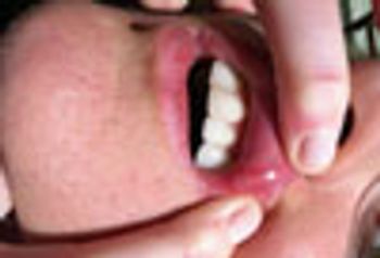
The shallow ulceration surrounded by a ring of erythema is characteristic for aphthous ulcers (canker sores). This patient’s otherwise negative history rules out Behcet’s disease.