
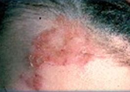


The 89-year-old has used topical mupirocin twice daily for several weeks without improvement. Examine the images. What's your diagnosis?

The 89-year-old has used topical mupirocin twice daily for several weeks without improvement. Examine the images. What's your diagnosis?
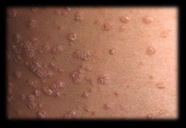
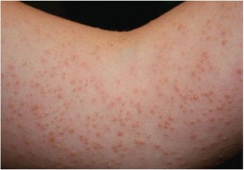
A summer tan failed to conceal this embarrassing rash on an 18 y/o boy’s arms, back, and chest. Can you make a diagnosis?

The young man is missing school because he is self-conscious about the hypopigmented lesions on his arms, back, and chest. Can you make the diagnosis?
Sonidegib becomes second hedgehog inhibitor for locally advanced disease.

These slides summarize the latest sun safety dos and don’ts, providing the most sound and sensible patient education recommendations under the sun.
The patient says the incidental finding appeared in adulthood and has been stable for years. Can you identify this reticulated erythema?
What are the characteristics of this blistering rash caused by exposure to UV light?
Is this pruritic lesion just unsightly, or is it indicative of a dangerous condition? Test your acumen in this quiz.
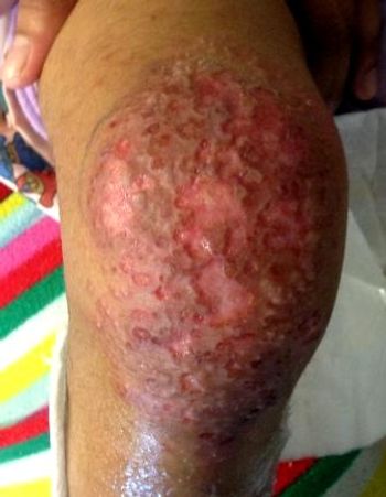
Two lesions have been slowly expanding on an older man’s chest for 2 years. What is the problem? This and questions on 3 other topics in this quiz.
This is a very distinct, rare, and remarkable hemorrhagic rash, first recognized in 2006, with 7 known cases reported in the literature.
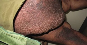
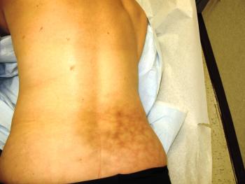
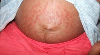
What’s the underlying cause of current cellulitis in this morbidly obese man?
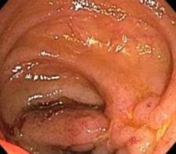
There is a lot of new information about the link between various malignancies and inflammatory bowel disease. Here: answers to 3 key questions.
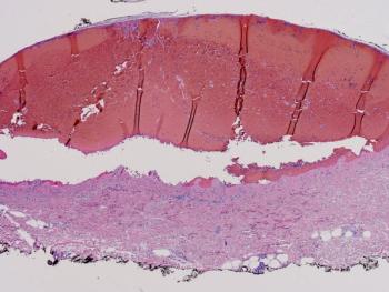
This diagnosis is a relative dermatologic emergency; presumptive treatment with antivirals should at least be considered if any suspicion exists.
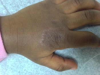
What figures into your differential of a slowly enlarging asymptomatic plaque, like the one on this child's hand?
A patient with a history of moderately severe and extensive plaque psoriasis now has a rash on her hands unlike any skin problem she has previously experienced. Do you know what this is? Answer this question and 4 others in this quiz.
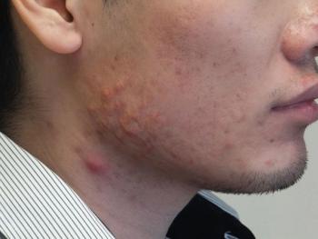
New guidelines recommend benzoyl peroxide for most patients with comedonal acne and all patients with inflammatory acne-especially those who receive a topical or oral antibiotic as therapy, in whom it can prevent antibiotic resistance.
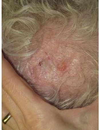
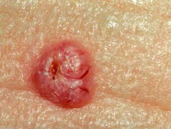
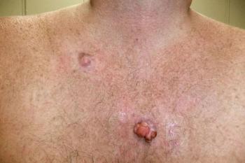
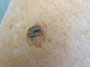
Would Mohs surgery be indicated for this 68-year-old man?
Texting without gloves while sitting on a ski lift explains the onset of this rash in a patient with a long history of widespread psoriasis.
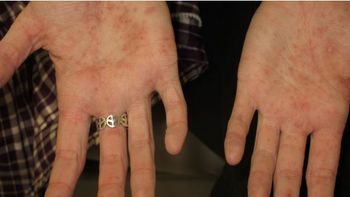
This woman was snowboarding over a very cold weekend and developed a pustular rash at night after her activities.
This lesion began to develop multiple colors. Is there cause for concern? Test your knowledge on 5 topics in this quiz.

This benign, solitary, nodular tumor on the lower eyelid arises from the follicular infundibulum. What do you know about this condition and the 4 others in this quiz?
This tongue-twister of a dermatosis is the most common type seen during pregnancy and primarily occurs in women carrying twins or triplets.
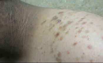
This 13-year-old boy presents with hyperpigmented skin on his neck. He is 66.75 inches and weighs 232 pounds. Your suspicion?

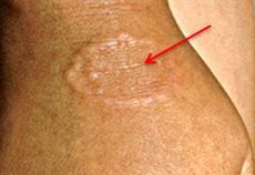
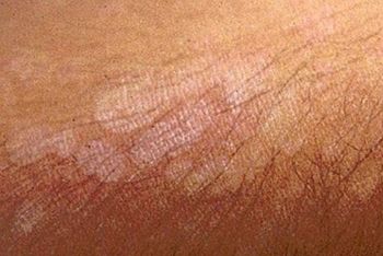
These discolored, scaly lesions on this young man’s trunk have recurred for the past 2 summers. He reports that the rash becomes “irritated and itchy” when he is in the sun.
You might not think of sarcoidosis in this patient’s case, but that’s the point. Here: teaching points about a disease not to be missed.