


A young teen presents with an acne outbreak that is uncharacteristic for her. What are the clues to the diagnosis in the PMH?
A young teen presents with an acne outbreak that is uncharacteristic for her. What are the clues to the diagnosis in the PMH?
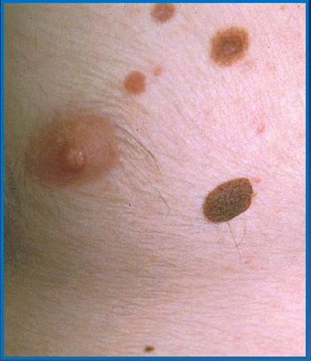
A young athlete presents with multiple moles on his chest and back. What’s a primary care physician to do? Test your knowledge with this quiz.
What's the matter with these kids? Get a close look at and a brief overview of seven common dermatoses seen in childhood.

What is in your differential diagnosis for a thin brown line of parallel bands, regular in width that span nail fold to free nail edge?
You will likely see these rashes in the course of primary care practice. Take this quick quiz to see what you know.
Would any of the 7 lumps and bumps here give you cause for alarm? Get an up-close look and a short lesson on each.
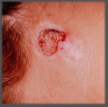
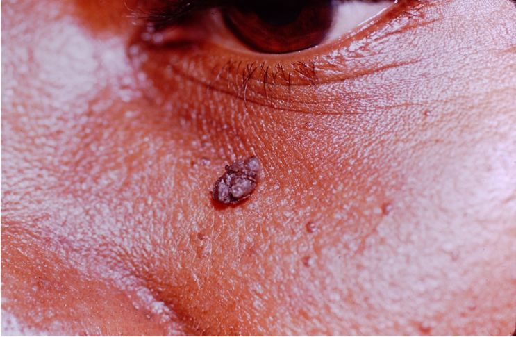
This lesion on the posterior neck of a young woman was present since birth and recurred after electrodessication. Can you Dx?
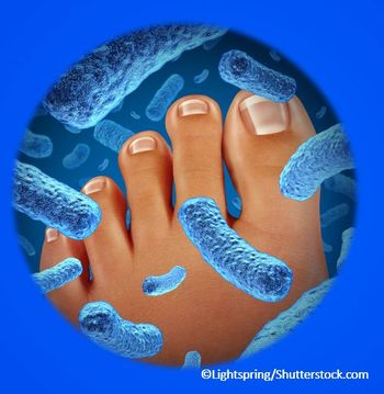
Take this quick quiz to test your knowledge of a common cutaneous bacterial infection.

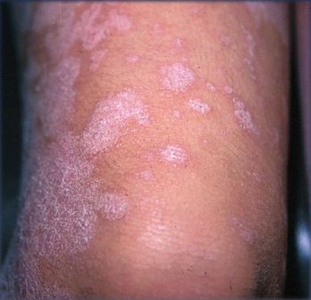
An acute outbreak on a formerly clear-complected young girl is suspicious for some notable features that are missing. Your Dx?
The causes are diverse, and the diagnosis may be challenging. Take this quick quiz to test your knowledge.
Disruption in the epidermal barrier creates susceptibility to bacterial and viral colonization that can range in severity from benign to dermatologic emergency.

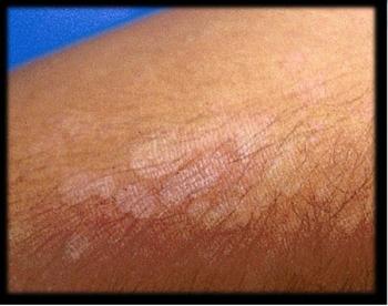
The first stage of this rash began at birth; it has twice changed distinctly in form. Are these changes the key to a diagnosis?
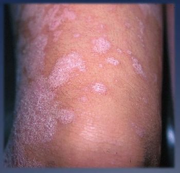
Diagnosis and treatment of tinea infections can be difficult. Take this quick quiz to test your knowledge.
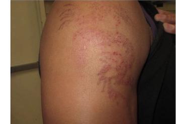
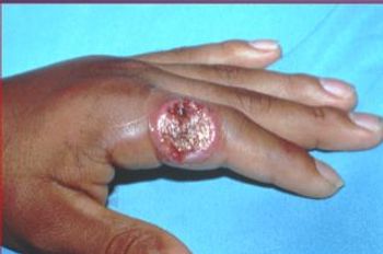
When in the Caribbean, why not get a tattoo? This man could now tell you exactly why to resist the vacation temptation.
Take this quick quiz to test your knowledge of a relatively uncommon inflammatory muscle disease.

Can you identify this acute widespread nodular rash on a neonate who is otherwise ready for discharge?
The condition is common but seen infrequently in primary care practice. Take this quiz for a quick refresher.
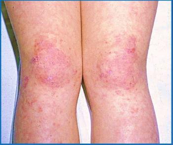
Lesions are warm to the touch; exam is unremarkable. Does mom have the answer? Do you?
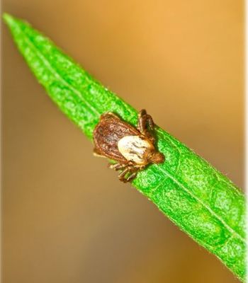
Correct diagnosis of Rocky Mountain spotted fever leads to correct treatment, but many cases are misdiagnosed. Take this test to see what you know.
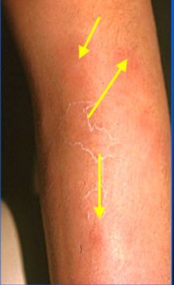
Skin ulcerations have a broad differential. Take this quick quiz to test your knowledge of some difficult diagnoses.
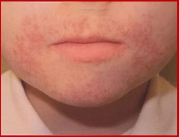
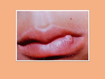
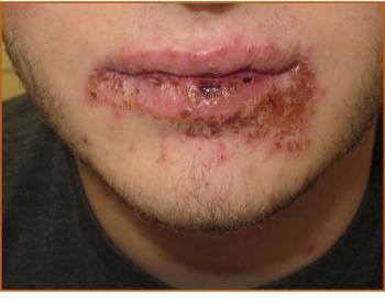
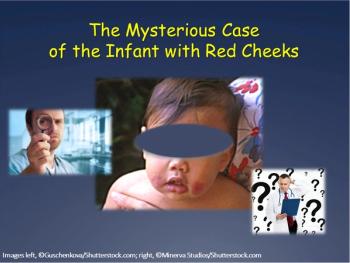
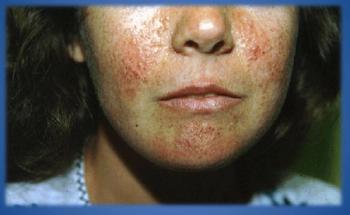
The eruption has been present for 1 year and is restricted to the perioral region. What is included in your differential?

Walk through this case of unexplained bruising in a young boy that proves signs and symptoms are not always what they appear to be.

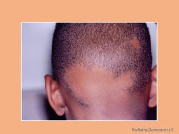
A boy’s bald spot has been present since birth. Take this quiz to test your knowledge of the most common congenital cicatricial alopecia.

What is the cause of this erythematous rash that appeared suddenly and spread rapidly on the child's trunk and extremities?
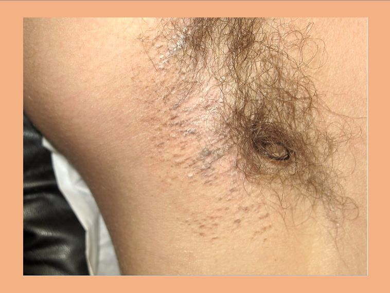
A young mother’s “high coloring” consists of papular, flesh-colored lesions that resemble telangiectasia. Can you ID?