


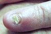
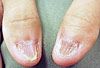


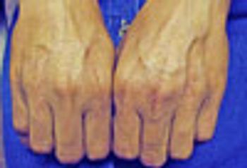
A 23-year-old man presented for medical attention with these mildly to moderately pruritic lesions located only on his hands. He was in good health and took no medications.
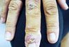
A 23-year-old man presented for medical attention with these mildly to moderately pruritic lesions located only on his hands. He was in good health and took no medications.
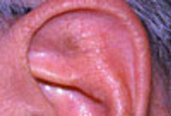
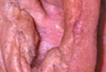
Skin lesions in the outer ear-the pinna, the concha, and the external auditory meatus-may be trivial or potentially malignant. Here we consider a flesh-colored swelling with a central white spicule; a tan papillary lesion; a fluctuant swelling; a red berry-like papule; an elevated pink lesion.
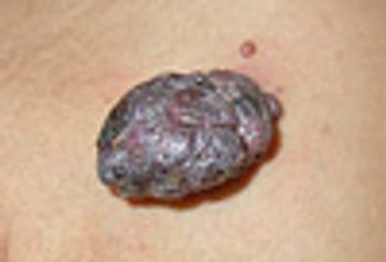
Benign skin lesions can arise in the outer ear as easily as in any other body part frequently exposed to the sun. Seborrheic keratosis may mimic malignant melanoma but is innocuous. Actinic keratosis is premalignant and should be excised, biopsied, and the site of excision monitored vigilantly.
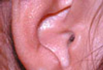
These innocuous lesions of the outer ear may arise spontaneously or after trauma or surgery. Both auricular seroma and pyogenic granuloma usually resolve satisfactorily after minor surgery, though they may recur.
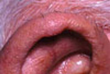
Diagnostic challenge: Two case reports of easily treated and innocuous causes of lesions in the outer ear. Chondrodermatitis nodularis helicis is associated with long cellphone use. Verruca vulgaris is caused, like all other warts, by human papillomavirus.
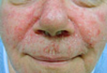
A 58-year-old man presents for evaluation of a facial rash. He is convinced, from internet research, that he has systemic lupus erythematosus.
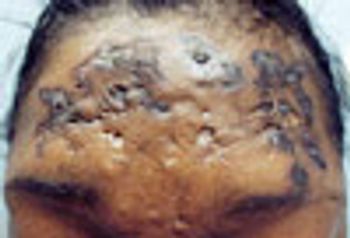
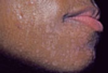
A 26-year-old African American woman was concerned about the gradual onset of mildly tender, but severely distressing facial lesions located on the face.
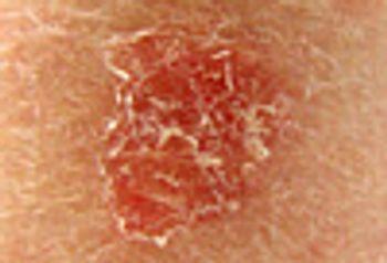
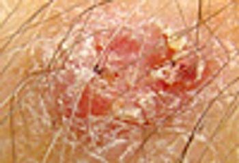
With no history of papulosquamous skin disease (eg, psoriasis), a solitary, well-demarcated, bright red scaly patch should suggest either superficial basal cell or in situ squamous cell carcinoma.
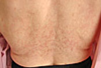
The husband of this 50-year-old obese woman noted that her back was discolored. Review of past medical history disclosed long-standing low back pain from partially herniated vertebral disks.
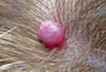
A 36-year-old woman notes the insidious onset of an asymptomatic lesion located on the scalp. Despite frequently hitting it with a comb or brush, it has never bled.
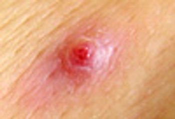
A 53-year-old woman felt the underwire in her bra stab the inframammary skin. She covered the ensuing shallow wound with antibiotic ointment under a bandage. The wound was slow to heal, and was ultimately replaced by a friable, oozing papule.
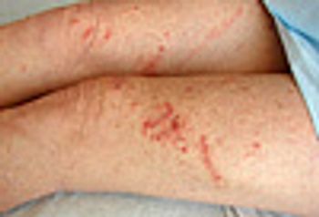
A 30-year-old woman rapidly developed an intensely pruritic eruption on both legs, that extended from the knees to the upper thighs.
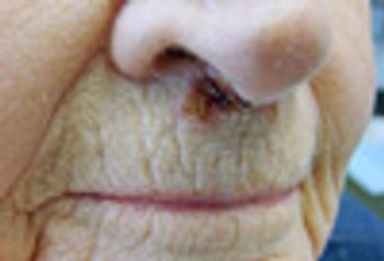
An 82-year-old woman presents with a persistent “face sore,” which she ascribed to repeatedly and frequently blowing her nose during a recent upper respiratory infection.
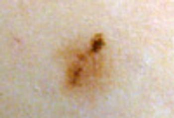
A 32-year-old woman sought medical attention after noticing a change in the appearance of a mole on her leg.
A 26-year-old man cut the dorsum of his wrist on a coral formation while snorkeling in the Caribbean.
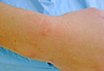
A 61-year-old obese, man with type 2 diabetes presented with the gradual onset of a mildly tender lesion on the dorsum of the right arm.
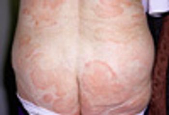
A 66-year-old woman is concerned about a gradually increasing rash located on the buttocks, lower back, abdomen, and upper, posterior thighs. The eruption has been present at least 18 months and is mildly pruritic.
A 36-year-old presents for evaluation of a rapidly enlarging nodule on the shoulder.
A 27-year-old woman asks about slightly itchy and scaly “white spots” on her face. She has rather severe dandruff, which requires shampooing every other day for control.
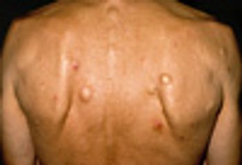
A 50-year-old man wonders whether the many lesions he has had for “almost all my life” can be removed. The lesions are asymptomatic, but cosmetically disagreeable to his third wife.
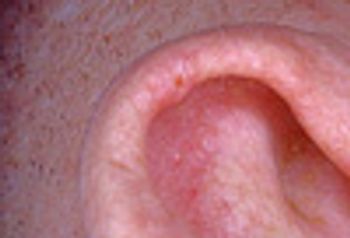
A 40-year-old man complains about a severely painful bump on his ear. He has trouble sleeping on that side due to the pain that occurs when he lays his head (and ear) down on the pillow. What is his condition?
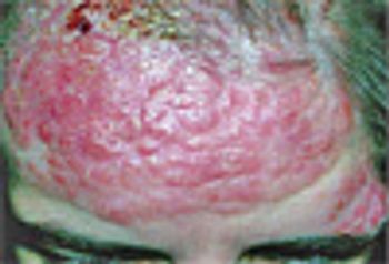
A 31-year-old woman presents for evaluation of an asymptomatic, firm plaque on the forehead. Lesions of similar size and consistency were present at the nape of the neck and on the chest. The patient was in overall good health. A biopsy performed to rule out cutaneous lymphoma revealed almost pure sheets of plasma cells in the dermis. What are your diagnostic suspicions and what would your next step be? Please click here and add your comments.
A 31-year-old woman presents for evaluation of an asymptomatic, firm plaque on the forehead. Lesions of similar size and consistency were present at the nape of the neck and on the chest.
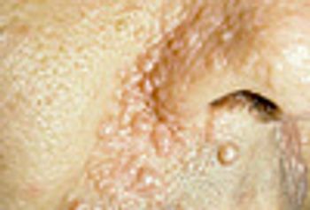
A 48-year-old man presents for treatment of herpes zoster. As an incidental finding, numerous flesh-colored, asymptomatic papules are noted on the central face.
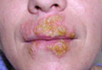
A 32-year-old woman presents with recurrent episodes of lip swelling associated with massive, painful blister formation and crusting.