


Although penicillin has been the antibiotic of choice for group A streptococcal (GAS) tonsillopharyngitis, a number of other antibiotics are used as well. This includes azithromycin, which may be preferred by some because of its dosing schedule. The results of a meta-analysis by Casey and Pichichero indicate that a total treatment dose of 60 mg/kg of azithromycin is necessary to eradicate GAS tonsillopharyngitis in children, and this regimen appears to be more effective than regimens of other antibiotics studied.
Infectious Disease
Latest News
Advertisement
Advertisement
Abstract: Although cystic fibrosis (CF) is typically diagnosed during infancy or childhood, it may escape detection until adulthood. Diagnostic accuracy can be sharpened by maintaining a high index of suspicion for CF in an adult who is pancreatic-sufficient but has unexplained recurrent respiratory infections, bronchiectasis, or nutritional deficiencies. The workup begins with the quantitative pilocarpine iontophoresis sweat test. If necessary, additional tests include mutation analysis, full-gene sequencing of CF transmembrane conductance regulator protein, and measurement of nasal transepithelial potential difference. Multidisciplinary care is essential and includes nutritional support, chest physiotherapy, exercise, appropriate antibiotics, and other pulmonary interventions. Dornase alpha, inhaled tobramycin, and azithromycin have been associated with improved outcomes and are considered to be the standard of care for patients with moderate lung involvement. (J Respir Dis. 2006;27(1):32-41)
The influenza vaccine has been used for many years to control outbreaks of influenza, and its role in reducing morbidity and mortality is widely appreciated among health care professionals and patients alike. The panic that occurred in 2004 after announcements of a vaccine shortage bears testimony to the importance placed on this approach to influenza prevention and control.
Abstract: Shortness of breath is a common complaint associated with a number of conditions. Although the results of the history and physical examination, chest radiography, and spirometry frequently identify the diagnosis, dyspnea that remains unexplained after the initial evaluation can be problematic. A stepwise approach that focuses further testing on the most likely diagnoses is most effective in younger patients. Early bronchoprovocation challenge testing is warranted in younger patients because of the high prevalence of asthma in this population. Older patients require more complete evaluation because of their increased risk of multiple cardiopulmonary abnormalities. For patients who have multiple contributing factors or no clear diagnosis, cardiopulmonary exercise testing can help prioritize treatment and focus further evaluation. (J Respir Dis. 2006;27(1):10-24)
The patient feels well and denies any symptoms; specifically, he has no pruritus, fever, weakness, fatigue, GI symptoms, or cardiac symptoms.
ABSTRACT: A scheme-based approach, supported by a simple mnemonic, can narrow the broad differential diagnosis of thrombocytopenia. This approach uses findings from the complete blood cell count and the peripheral smear to organize the possible causes of thrombocytopenia into those that affect only platelet count, those that produce both a low platelet count and hemolytic anemia, and those that produce disturbances in all 3 blood cell lines. Causes of isolated thrombocytopenia include viral infections, immune-mediated platelet destruction, congenital diseases, gestational thrombocytopenia, conditions in which splenomegaly is a prominent feature, antiphospholipid antibody syndrome, infectious diseases of bacterial origin, and drugs. Causes of thrombocytopenia in conjunction with hemolytic anemia include hemolytic uremic syndrome, thrombotic thrombocytopenia purpura, and disseminated intravascular coagulation. Disorders that produce disturbances in all 3 blood cell lines include aplastic anemia, myeloproliferative syndromes, myelodysplasia (both primary and secondary), myelofibrosis, myelophthisis, and several other diseases in which splenomegaly is prominent.
16-month-old previously healthy child is hospitalized after 36 hours of worsening painful edema and erythema of the right lower leg and high fever with chills.
ABSTRACT: A number of nondental conditions may cause significant oral pain. Pain associated with temporal arteritis is localized to the maxillary posterior teeth, the maxilla, or the frontal-temple region. This pain is often associated with exquisite tenderness of the scalp and face. The pain of trigeminal neuralgia is typically felt in the anterior maxillary or mandibular anterior teeth; it radiates along the mandible toward or into the ear on the ipsilateral side of the trigger. Pain may remit for months or years but is often severe when it recurs. Burning mouth syndrome preferentially affects postmenopausal women older than 50 years; one half to two thirds of patients experience spontaneous remission within 6 to 7 years, with or without treatment. The pain of postherpetic neuralgia is unilateral and restricted to the affected dermatome; it may be aggravated by mechanical contact or chewing.
A 56-year-old woman presents for a routine examination. She has been healthy, and results of previous examinations have been normal.
Erythema and bulla formation characterize the typical lesions of superficial frostbite. Deep frostbite, in contrast, involves subcutaneous tissue and usually leads to tissue loss.
For 2 months, a 19-year-old woman has had a slightly tender acneiform eruption on the trunk that spread to the extremities.
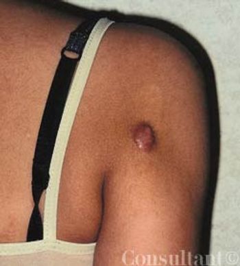
An 18-year-old girl presented with an asymptomatic nodule on the posterior aspect of the right upper arm. The lesion had developed a month after an episode of chickenpox at 11 years of age and had slowly enlarged. The lesion was 7 mm in diameter; it was firm, rubbery, reddish brown, and nontender.
A 45-year-old man was referred to our pulmonary clinic for progressive dyspnea and worsening asthma. His shortness of breath had been worsening over the past 2 years. He denied fever, weight loss, and other systemic complaints.
In the October 2004 issue of TheJournal of Respiratory Diseases,Morrison and Gupta1 reviewed theclinical and laboratory approachesto the diagnosis of communityacquiredpneumonia (CAP) causedby Legionella. They discussed theadvantages and limitations of culture,direct fluorescent antibody(DFA) staining, serology, polymerasechain reaction, and Legionellaurinary antigen assays. As the authorsnoted, DFA staining of respiratorysecretions is an underusedtest that has a high specificity in patientswith untreated Legionnairesdisease.
Scott and associates described a case of Phialemonium obovatum fungemia in a bone marrow transplant recipient. This fungal infection was associated with caseating granulomata in the lungs and GI involvement that resembled Crohn disease.
Although lipoprotein levels are known to be reduced in critically ill patients, the prognostic significance of this in patients with sepsis has not been established. However, a study recently conducted in Taiwan is worth noting; it found that low levels of high-density lipoprotein (HDL) cholesterol on day 1 of severe sepsis were associated with increased risk of death.
Abstract: The manifestations of indoor mold-related disease (IMRD) include irritant effects, such as conjunctivitis and rhinitis; nonspecific respiratory complaints, such as cough and wheeze; hypersensitivity pneumonitis; allergic fungal sinusitis; and mycotoxicosis. The diagnosis of IMRD depends on eliciting an accurate history and excluding preexisting pathology that would account for the patient's symptoms. Laboratory tests, imaging studies, and spirometry can play an important role in ruling out other diagnoses, such as allergic or nonallergic rhinitis, asthma, and pneumonia. The diagnosis of IMRD also involves integrating the results of immunologic, physiologic, and imaging studies with the results of indoor air-quality studies. (J Respir Dis. 2005;26(12):520-525)
Abstract: Spinal tuberculosis is the most common form of osteoarticular involvement in patients with tuberculosis. Localized pain is a common presenting symptom. In patients who do not present until vertebral wedging and collapse have occurred, a localized knuckle kyphosis is obvious, especially in the dorsal spine. In some patients, a retropharyngeal abscess develops, causing dysphagia, dyspnea, and/or hoarseness. Peripheral joint tuberculosis is characterized by an insidious onset of slowly progressive, painful, and swollen monoarthropathy, most commonly affecting the hip or knee. The radiologic features include juxta-articular osteoporosis, peripheral osseous erosion, and gradual narrowing of the interosseous space. Treatment involves antituberculosis drugs; the indications for surgery are relatively limited. (J Respir Dis. 2005; 26(12):543-546)
A 25-year-old man presented to the emergency department with left scrotal swelling. He mentioned that he also had severe intermittent right-sided chest pain of 1 week's duration that began while he was lifting heavy items at work. The pain worsened with exertion and was relieved by rest. There was no radiation to the shoulder or back. The patient's medical history was otherwise not significant.
Abstract: Acute chest syndrome (ACS) is one of the most common causes of death and hospitalization among patients with a sickle hemoglobinopathy. The clinical presentation is characterized by the appearance of a new infiltrate on a chest radiograph, with 1 or more new symptoms, including fever, cough, chest pain, and dyspnea. Additional findings include leukocytosis, hypoxemia, and auscultatory signs of consolidation. The differential diagnosis includes pneumonia, pulmonary infarction, fat embolism syndrome, pulmonary edema, and bone infarction. Treatment of ACS involves supportive care, empiric antibiotic therapy, and red blood cell transfusion when indicated. The decision of whether to use simple or exchange transfusions depends on the severity of illness and the risk of acute respiratory failure. Currently, hydroxyurea is the only FDA-approved drug designated as a preventive therapy. (J Respir Dis. 2005;26(12):529-534)
A 16-year-old boy visiting his aunt and uncle at a cabin in the country was bitten by their cat earlier in the day. He tried to pick up the animal after it had been injured, and it bit him on the dorsum of the hand between the thumb and first finger.
A 57-year-old woman complains of burning and dryness in her left eye and altered sensation in her mouth when eating; these symptoms began the day before. A coworker who had noticed facial asymmetry recommended that she seek medical attention.
I am concerned about Dr Benjamin Barankin's recent "Photoclinic" case of an alleged brown recluse spider bite.
A 49-year-old man has had dyspnea on exertion for 1 month; it has worsened during the past 2 days. He has also had 2 episodes of epistaxis and increasing abdominal distention, without pain, during the past 6 months.
Chronic diarrhea presents difficulties for clinicians as well as for patients. Because the differential diagnosis is enormous, management can be challenging. In this article, we present a strategy for quickly narrowing the differential based on a simple analysis of stool characteristics. We then describe an appropriate workup for each of the basic types of diarrhea.
Advertisement
Advertisement
Trending on Patient Care Online
1
Newer GLP-1 Therapies Linked to Fewer Alcohol-Related Hospitalizations in Adults With AUD
2
Early Liver Disease Detection Starts in Primary Care: A Q&A With Stevan Gonzalez, MD
3
Compulsive Smartphone Use Linked to Depressive Symptoms in Older Adults
4
2026 Dyslipidemia Guideline Expands Statin Eligibility to 21.5 Million More US Adults
5