


Extreme synovial inflammation resulted in the articular degradation seen in the hands of this 40-year-old woman who has Still disease, or juvenile rheumatoid arthritis (JRA).
Infectious Disease
Latest News
Advertisement
Advertisement
ABSTRACT: The basic screening studies for rheumatic diseases are a complete blood cell count, a determination of the erythrocyte sedimentation rate (ESR) or C-reactive protein (CRP) level, a rheumatoid factor assay, an antinuclear antibody (ANA) test, a measurement of serum uric acid level, and a urinalysis. Test results must be interpreted within a clinical context; for example, a positive ANA assay suggests the possibility of a rheumatic disorder, but it is not specific for any diagnosis. Tests that reveal the nature and extent of target-organ involvement, such as renal function studies in patients with systemic lupus erythematosus, can help guide the selection of therapy. Laboratory results also reflect disease activity; the ESR and CRP level are useful gauges of the activity of most inflammatory rheumatic disorders. Finally, laboratory monitoring can help you minimize the significant toxicity associated with many of the drugs used to manage rheumatic diseases.

A52-year-old white man presented with a pruritic eruption on the neck of 3 months’ duration. The rash had not responded to a potent topical corticosteroid prescribed by another practitioner for the presumed diagnosis of eczema. The patient reported no current health problems. His history included a pubic louse infestation and several episodes of uncomplicated urethral gonorrhea. He readily admitted to having unprotected sexual intercourse with prostitutes.
A 24-year-old man presented for evaluation of pruritic vesicles on both feet.Ten days earlier, dyshidrotic eczema had been diagnosed by another physicianwho prescribed triamcinolone ointment. The patient reported that the footeruption worsened after the topical medication was applied.
A 70-year-old man first noticed thisskin condition when he returned fromthe South Pacific at the end of WorldWar II. Over the years, the rash hasitched only occasionally; however,during a recent spate of hot weather,the eruption became highly pruritic.Applications of an over-the-counter1% hydrocortisone ointment exacerbatedthe condition
An eruption on the face of a 49-year-old woman had been misdiagnosed as astaphylococcal infection; the rash failed to respond to oral and topical antibiotics.A mid-potency topical corticosteroid also had been tried, but the eruptionworsened.
The parents of a 3-year-old girl sought evaluation of their daughter’s hair loss.During the past several months, a large patch of alopecia with scaling had developed.The differential diagnosis included seborrhea, trichotillomania, andtinea capitis.
A new lesion recently arose on the right flexor forearm ofa 67-year-old man. The 1-cm, pruritic, pink, circular, slightlyraised lesion was perfectly homogeneous with no centralclearing.
A 49-year-old man was concerned about a right flexor forearmlesion that had been increasing in size for 6 weeks.The light pink, well-demarcated, 5-cm, circular lesion featuredslight peripheral elevation with ulceration, crusting,and a relatively clear central area. A culture of materialfrom the lesion was negative for fungi. A potassium hydroxideevaluation was not performed.
A 56-year-old man had an asymmetric,maculopapular, sharply demarcated,pruritic, excoriated dermatitis on hisupper thighs. The eruption had beenpresent for 2 to 3 weeks
For about 4 months, a very dry, diffuse,fine scaly, asymptomatic eruptioncovered the palms of a 28-yearoldman; several fingernails weredystrophic bilaterally as well. Beforethe onset of this condition, bilateralonychomycosis of the toenails hadbeen diagnosed. The toenails had notbeen treated and were still affectedat the time of presentation. Branchinghyphae were seen on a potassiumhydroxide preparation of a fingernailcutting. The patient had tinea manuumand tinea unguium
Numerous factors contribute to the medication errors that kill up to 98,000 patients each year. Unnecessarily high dosages can result in increased side effects with only a small therapeutic benefit, especially in elderly patients. Lack of patient information-such as a history of allergies or adverse drug reactions-is another cause of error and injury. Communication failures include the use of ambiguous abbreviations, misinterpretation of verbal orders, and lack of timely response to a patient's medication-related symptoms. Dosing errors are common in children because of variability in dosage expressions in drug references. Remedies for prescribing errors are described in detail here.
A methodical approach to diagnosis usually reveals the cause of fever. In patients with simple fever, a careful history taking and physical examination combined with basic laboratory and imaging studies (complete blood cell count with differential, urinalysis, and possibly a chest film and blood cultures) usually yield the diagnosis. In patients with prolonged fever whose cause remains undiagnosed after extensive examination (fever of unknown origin), repeat the history taking and physical examination; also order routine laboratory studies, an HIV test, a tuberculin skin test, 3 sets of blood cultures, and chest films. In addition, abdominal CT scanning is often useful. Further testing at this point may include fluorodeoxyglucose positron emission tomography, technetium-tagged white blood cell scanning, transesophageal echocardiography, liver biopsy, bone marrow examination, and/or temporal artery biopsy. Exploratory laparotomy is rarely indicated.
Tinea infections can be diag- nosed by potassium hydroxide (KOH) examination, which reveals fungal elements when a preparation of scale from a lesion-particularly the active border-is studied under a microscope; culture; and histopathologic examination of a skin biopsy specimen or nail clippings with periodic acid–Schiff stain. Culture may be warranted when a fungal infection is strongly suspected despite a negative KOH result. Unfortunately, dermatophyte cultures can take from 4 to 6 weeks to become positive; therefore, treatment decisions may have to be made before culture findings are reported. A topical antifungal is the initial therapy for tinea cruris, tinea corporis, tinea pedis, and tinea manuum. Tinea capitis, extensive tinea corporis, and tinea unguium are best treated initially with oral antifungal agents, because these infections usually do not respond to topical therapy.
A 13-year-old girl of African American descent is brought to the pediatrician’s office becauseof a lesion on her neck. The girl’s mother had telephoned the office before the visit, statingthat the lesion resembled a blister at first, but now looked like a burn.
A 67-year-old Hispanic woman is seen for routine physical examination. Has mild hypertension but no other known medical problems. Feels well. No weight loss. No reported difficulty with eating, speaking, or swallowing. Denies any discomfiture in the mouth. States that nothing has changed in her mouth “ever since I lost my baby teeth.” Does not smoke cigarettes nor drink alcohol.
An 86-year-old woman had noted intermittent, transient “shading” and “hazing” of the vision in her right eye. Her best corrected vision in that eye was 20/20. She had pseudophakia from past cataract surgery.
An 87-year-old woman complained ofseeing a red tinge on the wallpaper inher house through her right eye. Thepatient had mild memory loss andmoderate hypertension, for whichshe took atenolol. She had quit smokingcigarettes many years earlier.
During an annual eye examination, a 65-year-old womanwith a 5-year history of type 2 insulin-dependent diabetescomplained that her vision had slightly worsened in botheyes. Her best corrected visual acuity was 20/30 in botheyes.
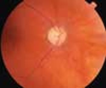
Sudden profound vision loss in her left eye prompted an82-year-old woman to seek evaluation. She also complainedof “just not feeling well” and reported new-onsettemporal and occipital headaches of 6 weeks’ duration.
A 49-year-old woman presented for aroutine eye examination. She had nohistory of systemic disease and tookno medications.
Colorectal cancer (CRC) is highly preventable; however, it remains a significant cause of morbidity and mortality in Western countries. CRC develops in more than 125,000 Americans each year, and about 50,000 die of it.1 Screening and early intervention significantly reduce morbidity and mortality, and a number of organizations have published screening recommendations (Table). Nevertheless, only 1 of every 3 eligible adults elects to be screened.2
Colorectal cancer (CRC) is highly preventable; however, it remains a significant cause of morbidity and mortality in Western countries. CRC develops in more than 125,000 Americans each year, and about 50,000 die of it.
Bronchodilators, preferably inhaled, are recommended for all patients with chronic obstructive pulmonary disease; ipratropium, with a 6- to 8-hour duration of action, is effective maintenance therapy. Tiotropium is currently being reviewed by the FDA for release in the United States; its once-daily dosing schedule may facilitate adherence. Criteria for long-term oxygen therapy are severe hypoxemia (PaO2, 55 mm Hg or lower) or a PaO2 of 60 mm Hg or lower with signs of cor pulmonale or secondary polycythemia (hematocrit higher than 55%). When symptoms are disabling despite optimal medical management, referral for pulmonary rehabilitation is the next step. Patients with upper lobe-predominant emphysema and low exercise capacity may benefit most from lung volume reduction surgery. Consider transplantation if the patient has severe lung disease that is refractory to medical therapy and survival is expected to be less than 2 to 3 years.
The key factor in reducing morbidity and mortality in patients with chronic obstructive pulmonary disease (COPD) continues to be smoking cessation. Newer formulations of nicotine replacement therapy-a nasal spray and an inhaler-provide rapid delivery of nicotine and may be appropriate for highly dependent smokers. Bupropion has been shown to improve smoking cessation rates, either when used alone or with a nicotine patch. Both the influenza and pneumococcal vaccines are recommended to reduce the morbidity and mortality associated with respiratory infections in patients with COPD.
Advertisement
Advertisement
Trending on Patient Care Online
1
FDA Clears First Generic Baloxavir for Influenza Treatment, Postexposure Prophylaxis
2
FDA Clears OTC Adapalene-Benzoyl Peroxide Gel for Acne in Patients 12 and Older
3
7 Drugs Approved for Primary Care: Q2 2026
4
How Primary Care Can Close DKA Monitoring Gaps Before Hospitalization
5