


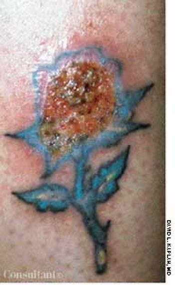
A 32-year-old woman with insulin-dependent diabetes noted a painful erosion at the site of "the rose tattoo," which she had gotten 5 days before.
A 32-year-old woman with insulin-dependent diabetes noted a painful erosion at the site of "the rose tattoo," which she had gotten 5 days before.
Abstract: Tuberculous meningitis has several different clinical presentations, including an acute meningitic syndrome simulating pyogenic meningitis, status epilepticus, stroke syndrome, and movement disorders. Cranial nerve palsies and seizures occur in about one third of patients, and vision loss is reported by almost 50%. The cerebrospinal fluid (CSF) typically shows moderately elevated levels of lymphocytes and protein and low levels of glucose. The demonstration of acid-fast bacilli in the CSF smear or Mycobacterium tuberculosis in culture confirms the diagnosis. CNS tuberculosis may also manifest as intracranial tuberculomas. The characteristic CT and MRI finding is a nodular enhancing lesion with a central hypointensity. Antituberculosis treatment should be initiated promptly when either tuberculous meningitis or tuberculoma is suspected. (J Respir Dis. 2005;26(9):392-400)
A 55-year-old man complains of fatigue. Although he sleeps 8 hours every night, he has to push himself to perform his usual daily activities. He has also experienced loss of libido and episodic impotence, which he ascribes to the fatigue.
Scaling patches that resist antifungals; an outbreak of red papules; a velvety,hyperpigmented rash--can you identify the disorders pictured here?
ABSTRACT: The management of chronic daily headache is difficult and complex. Those affected have a sensitive nervous system, and their predisposition for a low tolerance to sensory stimuli appears to be inherited. Under appropriate conditions, the equilibrium or balance between bombardment from painful stimuli and the regulatory systems that inhibit those stimuli is disrupted, allowing painful stimuli to become manifest at a greater intensity than in the nonmigraineur. Successful management depends on close adherence to nonpharmacologic approaches and pharmacologic regimens that desensitize the system and restore equilibrium. Comorbid conditions must be identified and treated as well.
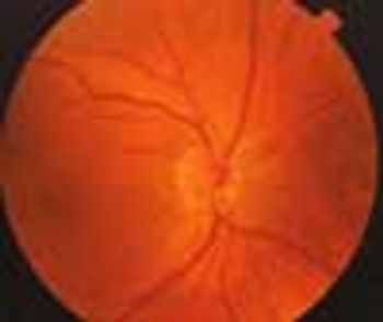
A 62-year-old man was referred for consultation by his optometrist because of a suspicious-looking optic nerve head in the left eye. The patient felt that his vision was good, and he had no history of unusual eye findings. This was his first dilated eye examination.

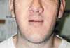
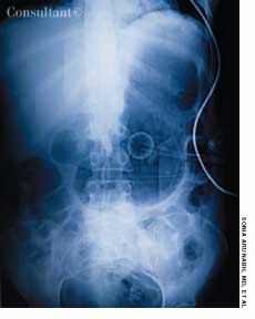
A 72-year-old woman first noticed progressive enlargement of the maxillary area of her face 18 years earlier. She denied facial trauma and significant dental caries. Her medical history consisted of breast cancer managed by a mastectomy and type 2 diabetes mellitus of 3 years’ duration.
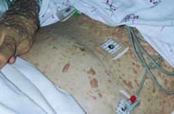
A 67-year-old woman with insulin-dependent diabetesmellitus and uncontrolled hyperglycemia complained offatigue and malaise. For 2 years, a draining ulcer hadbeen present on the bottom of her left foot.
ABSTRACT: The complexities of chronic nonmalignant pain and the contributions of psychosocial and environmental factors require assessment and treatment strategies that may go far beyond the use of analgesics. The first step is to rule out serious underlying conditions; determine the chronicity, quality, location, and intensity of the pain; assess aggravating environmental factors; and evaluate the patient's level of functioning. The pharmacologic regimen may include oral or topical analgesics, antidepressants, muscle relaxants, nerve stabilizers, and/or opioids. A comprehensive plan includes alternative modalities, such as physical and occupational therapy, stress management, relaxation techniques, and the treatment of comorbid conditions, including anxiety, depression, and sleep disorders. The optimal opioid regimen consists of methadone or a sustained-release opioid combined with a short-acting opioid for breakthrough pain.
A 23-year-old woman presents withweight loss, epigastric pain, abdominalfullness, and mild nausea. Shereports that she has had a slow-growingmass on her upper middle abdomen.She denies vomiting and doesnot have evidence of jaundice. Theonly significant finding in her medicalhistory is a myringotomy performedmany years earlier. She currentlytakes an oral contraceptive.
An 83-year-old woman is brought by her daughter for evaluation becauseof increasing confusion during the past few days. The patienthas early Alzheimer dementia, hypertension, and type 2 diabetes. She takes donepezil, 10 mg/d;lisinopril, 5 mg/d; and glipizide, 5 mg bid. She is unable to bathe and dress herself as well as previously,has been crying for no apparent reason, and has lost her appetite.
ABSTRACT: During the history taking, pay particular attention to pharmacologic and toxic exposures; among the medications and substances associated with tremor are ß-adrenergic agonists, stimulants, psychotropic agents, anticonvulsants, dopamine agonists, methylxanthine, and heavy metals. During the examination, observe the amplitude, frequency, and rhythm of the tremor. In contrast to physiologic tremors, which have low amplitude and high frequency, pathologic tremors typically have a higher amplitude and lower frequency. Tremors can be classified as resting, postural, or action. Resting tremor is almost always associated with other features of Parkinson disease, while postural and action tremors are prominent features of essential tremor. When essential tremor interferes with normal daily functioning, treatment is indicated. ß-Adrenergic antagonists, anticonvulsants, benzodiazepines, primidone, and nimodipine have been used in this setting. If these medications are ineffective, consider botulinum toxin A or surgery.
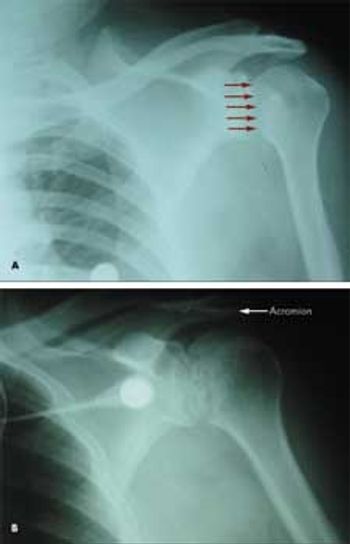
A 53-year-old man with type 2 diabetes mellitus and hypertension presented to the emergency department with pain in his left upper chest and back, neck, and shoulder. The pain increased with passive and active range of motion testing and decreased at rest. His physical examination was unremarkable except for restricted left shoulder movement and generalized tenderness in the left shoulder area.
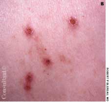
Since adolescence, a 67-year old woman had had multiple nodular lesions on her body that were painful at times, particularly when pressure was applied. She reported that other family members, including her mother and brother, had similar lesions.
We have recently had an upsurge in children with type 1 diabetes at our gastroenterologyclinic for celiac screening.
ABSTRACT: To assess a child for overweight, begin by calculating his or her body mass index (BMI). Note that BMI is used differently in children than it is in adults. A child's BMI is plotted on a growth curve that reflects that child's age and gender. This yields a value-BMI-for-age-that provides a consistent measure across age groups. Children whose BMI-for-age is between 85% and 95% are at risk for becoming overweight. Any child whose BMI-for-age is 95% or more is considered overweight. The 2 main factors associated with overweight in children are poor eating habits and decreased physical activity. Recommend that children have at least 5 servings of fruits and vegetables a day. Children should engage in moderate physical activity for at least 60 minutes on most days of the week, and TV viewing and computer activities should be limited to no more than 2 hours a day.
ABSTRACT: The Studies of Left Ventricular Dysfunction (SOLVD) trials demonstrated that early intervention in congestive heart failure (CHF) improves survival. However, early CHF is mainly a clinical diagnosis based on New York Heart Association criteria and, until recently, no easy and inexpensive screening test existed. There are now several such tests that employ radioimmunoassays (RIAs) to measure cardiac peptides in a single plasma sample; results help determine the likelihood that CHF is present but do not definitively establish the diagnosis. The vessel dilator RIA is the most specific and sensitive for differentiating persons with mild CHF from healthy ones; intravenous administration of this cardiac peptide hormone has beneficial hemodynamic, diuretic, and natriuretic properties in persons who have CHF. Brain natriuretic peptide (BNP) measured by fluorescence immunoassay is useful in the emergency department, because a result may be obtained in as little as 15 minutes. This assay may indicate CHF; further tests are recommended to define the diagnosis. BNP increases with other causes of dyspnea, including pulmonary hypertension, pulmonary emboli, and renal failure, so it is not specific for CHF. BNP also increases with age, and measured values are higher in women than in men.
A 60-year-old woman with hypertension, diabetes mellitus, and intermittentatrial fibrillation presents with nausea, diaphoresis, dizziness, and globalweakness that has lasted 1 hour. She denies chest pain, dyspnea, syncope,vomiting, diarrhea, blood loss, and headache; there is no vertigo. Medicationsinclude acetaminophen, digoxin, diltiazem, glipizide, hydrochlorothiazide,irbesartan, metformin, pioglitazone, and warfarin.
Swelling of the lower legs broughtthis 57-year-old woman to a familypractice clinic. She had a history ofhyperthyroidism with weight loss,tachycardia, and anxiety. This conditionwas confirmed with blood testsand radioactive iodine uptake testing.
This 17-year-old presented with a 1-month history of weight loss, increased appetite, mild insomnia, hand tremor, palpitations, sweating, heat intolerance, and quick loss of temper. The number of daily bowel movements had increased from 1 to 2. There was no family history of thyroid disorders.
Graves disease presents commonly with exophthalmos and stare and striking pretibialmyxedema.
Hyperpigmentation is seen on the cheeks and eyelids of a 36-year-old woman.She became hyperthyroid at age 19 years, with accompanying exophthalmosand hyperpigmentation, following the birth of her first child. Thyroidectomywas carried out at that time, and the patient has been receiving thyroid replacementtherapy ever since. The hyperpigmentation, an uncommon accompanimentof hyperthyroidism, has persisted.
Apidra (insulin glulisine injection)from Aventis is approved by the FDAfor use in adult patients with type 1 ortype 2 diabetes mellitus for the controlof hyperglycemia. Compared withhuman insulin, Apidra has a morerapid onset and a shorter duration ofaction. The drug is designed to managemealtime spikes in glucose levelsby administration through subcutaneousinjection or continuous subcutaneouspump infusion either 15 minutesbefore or 20 minutes after startinga meal.
ABSTRACT: The chief causes of ingrown toenails are trauma, incorrect toenail trimming, and tight footwear. Conservative measures, which include decreased activity, warm soaks, antibiotic therapy, elevating the edge of the impinged nail, debridement, or removal of the corner of the toenail containing the spike, may relieve moderate inflammation and infection. For patients who have severe impingement with acute infection, partial or complete nail avulsion is the treatment of choice. If avulsion is followed by recurrent infection, partial or complete ablation of the nail matrix may become necessary after the infectious process has resolved. Teach patients to trim their toenails straight across and not to curve them toward the lateral margins.
Over the previous 6 months, a59-year-old man had experienced lethargy,fatigue, poor appetite, cold intolerance,and abdominal distention. Hisvital signs were normal; physical examinationrevealed periorbital andpretibial edema, distant heart sounds,and delayed reflexes.