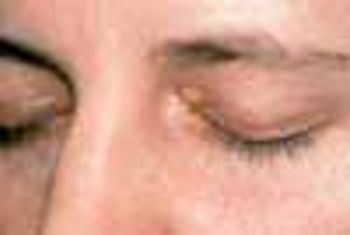
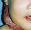
Here are two cases that demonstrate the discovery of aortic aneurysms through careful and complete physical examination and via radiographic studies obtained to evaluate other conditions.


Here are two cases that demonstrate the discovery of aortic aneurysms through careful and complete physical examination and via radiographic studies obtained to evaluate other conditions.

A 67-year-old woman presented to the emergency department (ED) with severe, sharp, central chest pain of sudden onset and mild shortness of breath. The pain had been present for 15 minutes. The patient was obese; her medical history included hypertension, myocardial infarction, and osteoarthritis.

A 54-year-old woman with a history of hypertension presented with a worsening headache and a left hemisensory defect. A CT scan of her head without contrast showed a right parietal hemorrhage with spreading edema; the masslike effect caused shifting of the midline to the contralateral side. The patient gradually became comatose and required intubation for airway protection. Intravenous corticosteroids were administered to decrease the effect of the lobar hemorrhage. Fever developed 3 days after admission.

Inflammation plays a major role in coronary artery disease (CAD), whereby inflammatory changes develop in the blood vessel walls.1 This observation has spurred interest in exploring the connection between CAD and markers of inflammation, including C-reactive protein (CRP), fibrinogen, serum amyloid A, and many other novel markers.

A 51-year-old man with a history of type B aortic dissection presented with severe right upper quadrant pain. He was febrile and hypotensive.

How much fish oil should I recommend that my patients with heart disease take?

Cardiovascular disease is a leading cause of death in patients with chronic obstructive pulmonary disease (COPD). While some physicians may be reluctant to prescribe ß-blockers for these patients, because of concern about adverse effects on lung function, a study conducted by Au and associates indicates that ß-blockers may have an edge over other antihypertensive agents in reducing mortality risk.

For 2 years, a 79-year-old man had postprandial fullness and epigastric discomfort. He also experienced regurgitation and substernal pain after eating that was relieved by belching. He had a history of hypertension and gout. The patient’s vital signs were normal.

The patient is a 47-year-old man who began to experiencefrequent headaches about 6 years before hepresented to a neurology clinic. The headaches rapidly progressedto become daily and almost constant. He describeda sensation of dull pressure in both temples that was presenton or within a few hours of awakening and that persistedfor the remainder of the day. He experienced a moreintense, disabling, throbbing pain in the same locationonce or twice a week, with photophobia and nausea, thatmight last 2 to 3 days. The patient took 2 to 6 over-thecounter(OTC) analgesic tablets each day-usually200 mg of ibuprofen. These would dull but not terminatethe pain.

A 67-year-old woman with insulin-dependent diabetesmellitus and uncontrolled hyperglycemia complained offatigue and malaise. For 2 years, a draining ulcer hadbeen present on the bottom of her left foot.

High-grade fever, chills, fatigue, malaise, and anorexia developed in a 35-year-old man following subclavian catheterization because of chronic renal failure of unknown cause. The patient, who had long-standing diabetes mellitus, was admitted to the ICU with the diagnosis of possible sepsis. The next day, he was found to have a grade 2/6 systolic murmur compatible with tricuspid regurgitation. This was confirmed when a 4-chamber echocardiogram (A) revealed a large single piece of vegetation (2 arrows) lying on the tricuspid valve, flapping in and out of the right ventricle. In a 2-dimensional echocardiogram of the right atrium and right ventricle (B), 3 arrows point to the vegetation. (RV, right ventricle; LV, left ventricle; RA, right atrium; LA, left atrium; TV, tricuspid valve.)

An 83-year-old woman is brought by her daughter for evaluation becauseof increasing confusion during the past few days. The patienthas early Alzheimer dementia, hypertension, and type 2 diabetes. She takes donepezil, 10 mg/d;lisinopril, 5 mg/d; and glipizide, 5 mg bid. She is unable to bathe and dress herself as well as previously,has been crying for no apparent reason, and has lost her appetite.

A 20-year-old woman presents with a 3-week history of a pruritic, progressivelyenlarging erythematous lesion on one arm. She has a cat and recentlystarted horseback riding lessons. She is otherwise healthy and takes nomedication.

Despite the manydouble-blind,placebo-controlledtrials thathave demonstratedthe efficacy of statins inreducing the risk of cardiovascularevents, a largenumber of patients who aretreated with these drugsstill experience suchevents. This may be becausepatients who requireintensive lipid lowering arenot receiving adequatedosages of statins.

Two weeks earlier, this 66-year-old man had been hospitalized because of leftsided chest pain. However, cardiac evaluation revealed no abnormalities.

A 53-year-old man with type 2 diabetes mellitus and hypertension presented to the emergency department with pain in his left upper chest and back, neck, and shoulder. The pain increased with passive and active range of motion testing and decreased at rest. His physical examination was unremarkable except for restricted left shoulder movement and generalized tenderness in the left shoulder area.

A 30-year-old man presentedwith severe left flankpain radiating to his abdomenand gross hematuriaof 5 to 10 days’ duration.He also reported a 4- to 6-monthhistory of nausea with intermittentvomiting, anorexia, and progressiveweight loss. He took no medicationsand had no allergies.

A mildly painful, nonpruritic rash on the forearms and legs prompted a 42-year-old man to go to the emergency department. The patient noted the rashwhen he awoke that morning. He had had joint pain and fever for the past7 days and generalized malaise with chills that began about 3 days earlier.He had no significant medical history.

Hypertensive crises encompass a spectrum of clinical situations thathave in common elevated blood pressure (BP) and progressive or impendingtarget organ damage. Each year more than 500,000 Americans (about1% of all persons with hypertension in the United States) have a hypertensivecrisis. In large urban areas, 25% of visits to the medical section of any givenemergency department (ED) are attributable to a hypertensive crisis.

A 35-year-old woman (gravida 3, para 2) presented at 25 weeks’ gestation with a 24-hour history of continuous, pressure-like, retrosternal chest pain, radiating only to the back. The pain worsened when she lay down, took deep inspirations, or coughed; it diminished when she leaned forward. The patient had no significant medical history and was not taking any medications; she denied fever and illicit drug use.

An obese 61-year-old man who hadchronic obstructive pulmonary diseaseand sleep apnea heard a “pop”in his stomach while lifting a heavyweight; severe abdominal pain followed.He was short of breath thenext morning, and his physician empiricallyprescribed cephalexin.

A 70-year-old man with a history of peptic ulcer disease presented with a 1-dayhistory of epigastric pain. Abdominal examination revealed mild epigastrictenderness. A pneumoperitoneum was discovered on a chest film (A), and aleft decubitus chest film (B) confirmed this diagnosis.

A 70-year-old woman with no previousmedical problems had had progressivedyspnea and generalized weaknessfor the past several days. She washypotensive (73/31 mm Hg), tachycardic(120 beats per minute), andtachypneic (28 breaths per minute);oxygen saturation (room air) via pulseoximetry was 84%.

Over the previous 6 months, a59-year-old man had experienced lethargy,fatigue, poor appetite, cold intolerance,and abdominal distention. Hisvital signs were normal; physical examinationrevealed periorbital andpretibial edema, distant heart sounds,and delayed reflexes.

A 72-year-old man with a history of rheumatic heart disease presented with pulmonary congestion and syncope. Results of the cardiac examination suggested mitral stenosis and atrial fibrillation with a controlled ventricular response.