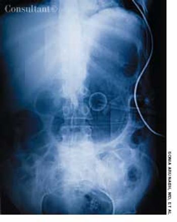
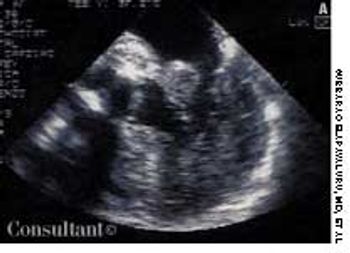
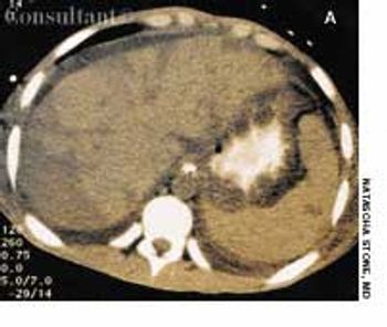
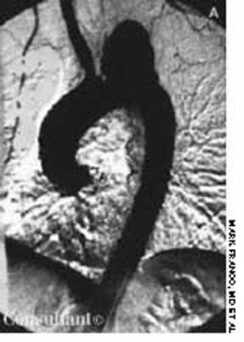
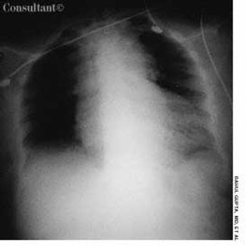
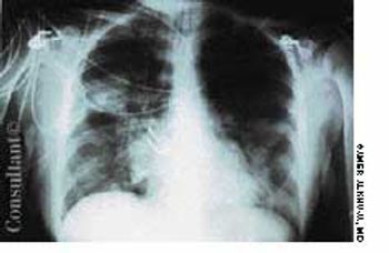
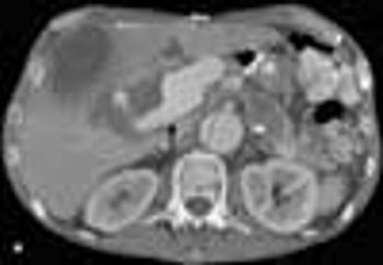
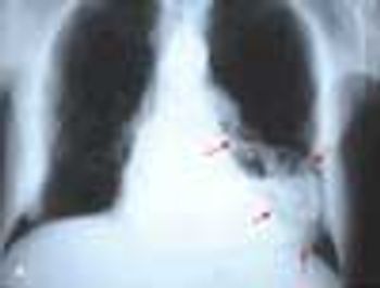
A 70-year-old man who had just completeda course of trimethoprim-sulfamethoxazolefor a urinary tract infectionpresented with palpable purpuraand cutaneous erosions of acute onseton his legs (A). He also had massivescrotal edema and purpura (B).

A 70-year-old man who had just completeda course of trimethoprim-sulfamethoxazolefor a urinary tract infectionpresented with palpable purpuraand cutaneous erosions of acute onseton his legs (A). He also had massivescrotal edema and purpura (B).

Painful blue toes developed in a 72-year-old woman with coronary artery andperipheral vascular disease after she underwent angiography.

A 16-year-old girl was bothered byankle pain and “red spots” on herlower legs. These symptoms clearedin a few days without treatment. Sixweeks later, after returning from anall-day outing at a fair, she noticedthat the spots had reappeared (A)and hemorrhagic lesions had developedon the right ankle (B) and leftheel (C). After removing her shoes,the teenager felt severe pain in bothankles, particularly the right.

A 57-year-old man with a history of venous stasis leg ulcerationwondered about the “white spots” on his leg. Thecondition is atrophie blanche, which manifests as smooth,ivory-white macules and plaques of sclerosis stippled withtelangiectasia that often are surrounded by mild to moderatepigmentation.

Redness, irritation, and diplopia developedover 2 to 3 weeks in a 55-yearoldman’s left eye (A). The injectionworsened and was unresponsive toeye drops. Ptosis, mild proptosis, andelevated intraocular pressure developed.A bruit was auscultated overthe affected eye.

Sudden pain and a pulsatile swellingof the right upper medial thigh concernedan 80-year-old woman (A).Coronary angioplasty had been performedthrough this site 5 weeksearlier.

Four months after a patchy, macular,erythematous spot erupted on thedorsum of a 63-year-old woman’s leftfoot, the area became ulcerated,tender, and painful. The 1.2-cm ulcerwas covered by a hemorrhagic crustsurrounded by a cyanotic reticulardiscoloration of the skin.

A 65-year-old woman, who was confined to a wheelchairbecause of severe rheumatoid arthritis, was concernedabout nodules that had erupted on her fingers and handsduring the previous 3 weeks (A). Her medical historyincluded colon cancer, chronic renal insufficiency, anemia,and hypertension. The nonpruritic nodules were painfulwhen they began to form under the skin; however, oncethey erupted, the pain disappeared.

Ten weeks before presentation, this55-year-old woman noticed decreasedsensation in her feet and a bluish discolorationof her toes. These symptomsprogressed rapidly, and pain andcoldness in both feet increased in intensity.Her feet subsequently becamegangrenous. Her seropositive arthritishad been diagnosed about 6 yearsearlier. The disease had been wellcontrolled until about 10 weeks beforethis photograph was taken.

Is hypertension a frequent manifestation of thyroid disease?Which clinical clues suggest hypertension in persons with thyroiddysfunction?

An 86-year-old woman had noted intermittent, transient “shading” and “hazing” of the vision in her right eye. Her best corrected vision in that eye was 20/20. She had pseudophakia from past cataract surgery.

An 87-year-old woman complained ofseeing a red tinge on the wallpaper inher house through her right eye. Thepatient had mild memory loss andmoderate hypertension, for whichshe took atenolol. She had quit smokingcigarettes many years earlier.

During an annual eye examination, a 65-year-old womanwith a 5-year history of type 2 insulin-dependent diabetescomplained that her vision had slightly worsened in botheyes. Her best corrected visual acuity was 20/30 in botheyes.

Sudden profound vision loss in her left eye prompted an82-year-old woman to seek evaluation. She also complainedof “just not feeling well” and reported new-onsettemporal and occipital headaches of 6 weeks’ duration.

A 49-year-old woman presented for aroutine eye examination. She had nohistory of systemic disease and tookno medications.

Q:Which combinations of antihypertensivemedications are most effective-and which arebest avoided?

Examine patients’ carotid arteries immediatelyafter you examine theirlungs.

Diffuse petechiae suddenly arose on the back and abdomen of a 79-year-old woman. Within several days, the asymptomatic lesions covered her arms and face as well.

A healthy 32-year-old scuba diver was taken to the emergency department (ED). He had lost consciousness following a dive in which he had held his breath during a rapid ascent to the surface and had performed the Valsalva maneuver to clear his ears.

A slightly pruritic, red, scaly rash on an 8-year-old boy’shands has been progressively worsening since it appeared4 months earlier. Nail pitting also was noted. There are noother rashes on his body. The patient is active in sports;denies any new exposure to soaps, clothing, or other contactants;and spends time in the homes of his recently divorcedparents.

An obese 55-year-old woman had intermittent dysuria and frequent urination for 4 days and fever, abdominal pain, and vomiting for 3 days. Her history included type 1 diabetes mellitus, hypertension, and angina; her medications included insulin, nifedipine, and nitroglycerin.

Pyoderma Gangrenosum belongs in the differential diagnosis of lower extremity vasculitis-like ulcerating lesions.

Twenty-four hours after a car accident, a 47-year-old man with chronic low back pain-which had been diagnosed years earlier as "arthritis" of the spine-presented with back and chest pain and requested an opioid analgesic. The patient had a history of alcoholism and illicit drug use. Tenderness of the thoracic area was noted.

A 59-year-old man, who was unable to walk, was brought to the emergency department with severe leg pain, worsening weakness, increasing fatigue, chills, and fever of 3 days' duration. The symptoms began after a round of golf. In addition to the leg pain, which particularly affected the calf muscles, the lower extremities were red and swollen.

A 77-year-old African American man with type 2 diabetes mellitus and coronary artery disease presented to the emergency department with acute scrotal swelling and pain. His testicles were erythematous with focal areas of necrosis and associated tissue destruction. Similar skin changes were apparent in the lower abdominal and inguinal regions.