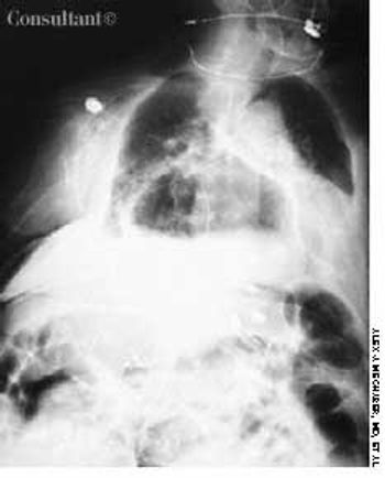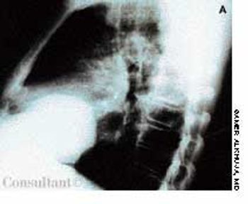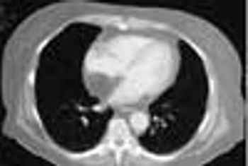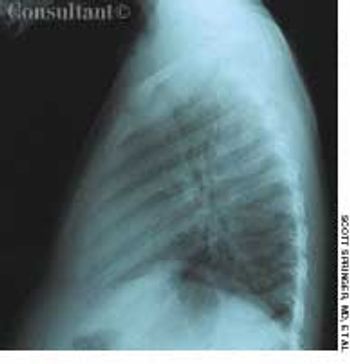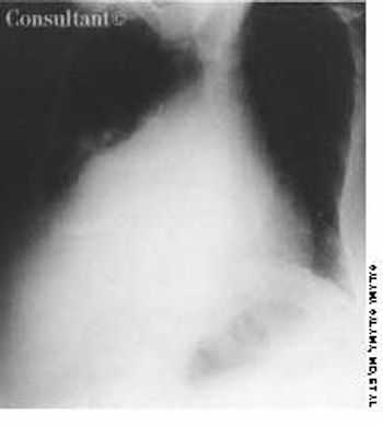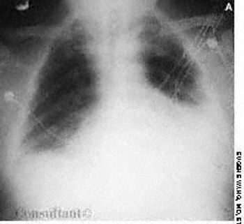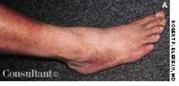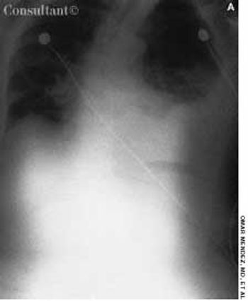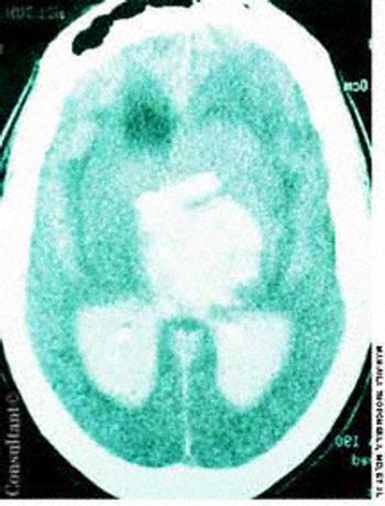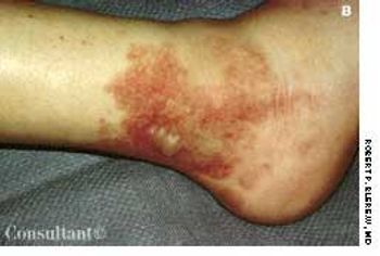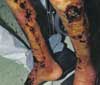

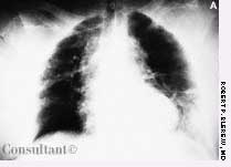
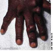


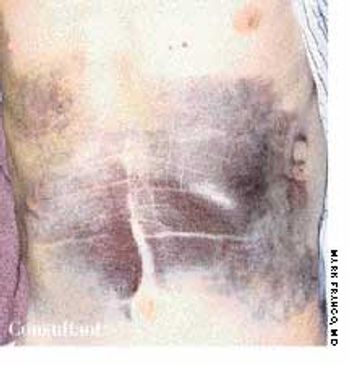
An 80-year-old man with a history of congestive heart failure, coronary artery disease, cardiomyopathy, and thoracic and abdominal aneurysms was taken to the emergency department because of mental status changes, back pain, and ecchymotic areas over his body. The ecchymoses started on his back 5 days before admission and spread to his abdomen.