


A 66-year-old woman was hospitalized because of severe anemia secondary to myelodysplastic syndrome. She had had associated fatigue and throbbing pain in both legs for several days.
Dermatology
Latest News
Advertisement
Advertisement
For 2 weeks, a previously healthy 40-year-old man has had excessive thirst and increased frequency of urination. He awakens at least 5 times every night to urinate. He reports no nausea, vomiting, change in bowel habits, chest pain, or dyspnea.
A 20-year-old white male collegiate basketball player has a 3-year history of marked facial and upper extremity flushing that occurs after about 20 minutes of indoor practice. The flushing is preceded by an intense sensation of coldness, and despite the very noticeable flushing, the involved areas are cold to the touch for some time.
Worsening painful ulcers on both legs prompted a 62-year-old woman to seek medical attention. She had a history of rheumatoid arthritis (RA), demonstrated by the markedly deformed interphalangeal joints in her thumbs (A), and scleroderma-polymyositis overlap syndrome.
A 74-year-old woman presented with a refractory pruritic eruption. Four months earlier, she had sought evaluation of a thickened, slightly crusted 6 3 8-cm patch on her right ankle of 2 months' duration. Contact dermatitis with secondary impetigo from scratching was suspected, and a topical corticosteroid and an oral antibiotic were prescribed.
A painful necrotic lesion, a pruritic rash that recurs after corticosteroid therapy, an ulcer on the tongue-do you recognize the disorders pictured here?
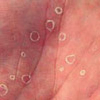
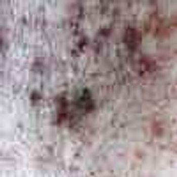
Six months after testing positive for HIV in 10 bands, a 24-year-old homosexual man presented with a macular rash on his palms and soles. He first noticed the lesions 2 weeks earlier; they were not pruritic or painful. He also had a brighter, more inflamed rash in the groin and antecubital fossae that was presumed to be a yeast infection and was treated with fluconazole. He had no other symptoms.
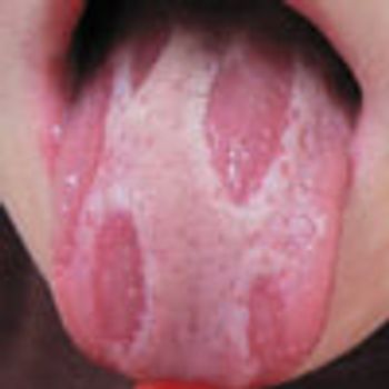
The sharply demarcated, smooth red plaques on this 3-year-old's tongue had been present for several months.
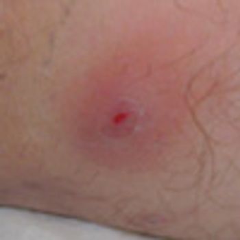
Hidradenitis suppurativa is a chronic acneiform infection of the cutaneous apocrine glands.
Within the past 7 years, the incidence of methicillin-resistant Staphylococcus aureus (MRSA) and Clostridium difficile infections has significantly increased. Risk factors for MRSA infection include previous antibiotic therapy and living arrangements such as prisons or military barracks that involve close, frequent contact with infected persons. Treat stable patients with MRSA skin infections with oral antibiotics in addition to incision and drainage; hospitalization and intravenous antibiotics are recommended for patients whose condition is unstable or who are unlikely to adhere to an oral regimen. A new strain of C difficile, BI/NAP1, has been associated with recurrent infection; more severe disease that mandates urgent colectomy; and dramatically higher mortality in vulnerable populations, such as older adults. Although oral metronidazole has been the mainstay of treatment of C difficile infection, oral vancomycin may be slightly more effective in patients with severe disease.
For the past month, a 67-year-old woman has had a pruritic rash under her left breast. She takes an antihypertensive and is otherwise healthy.
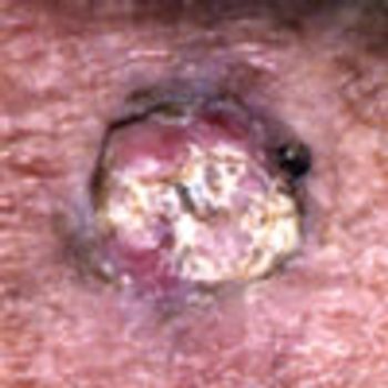
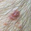
An 89-year-old man reported that this lesion began developing on his left forearm 11 days earlier. It is a keratoacanthoma, a rapidly growing but benign neoplasm that occurs predominantly on the extensor surfaces of the hands and forearms of white men over age 50.
For more than 3 years, a 63-year-old man with a long history of parapsoriasis had multiple hyperpigmented, erythematous plaqueswith scaling on the abdomen, back, feet, and arms. Some lesions had a hypopigmented center. The patient denied systemic symptoms.
This 8-year-old boy's mother thought her son had a fungal infection on his feet. Examination disclosed malodorous, nontender plaque formation on the weight-bearing surfaces of both feet. Within the plaques were round pits and furrows.
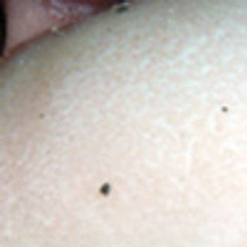
A 52-year-old man presented with asymptomatic papules on his scrotum. The lesions had first appeared 1 year earlier. He had not sustained local trauma to the scrotum, and his medical history was unremarkable. There was no family history of similar skin lesions.
A number of factors heighten the risk of parasitic infections in elderly persons, who often live in communal settings (eg, nursing homes), where they are more likely to encounter parasites, such as the scabies mite.
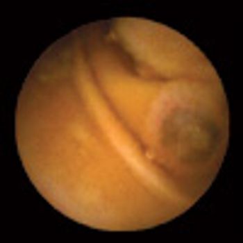
An 83-year-old man with a history of hypertension, hyperlipidemia, and diverticulosis was hospitalized because of painless hematochezia of 1 day's duration. Two years earlier, he had undergone surgical excision of a superficial spreading melanoma on his right thigh.
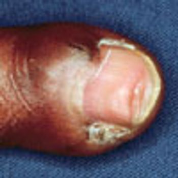
The left middle finger of this 30-year-old man was lacerated in a motorcycle accident. After it was surgically repaired, the finger developed some dystrophy as well as a small, separate fingernail in the lateral sulcus of the proximal nail fold.
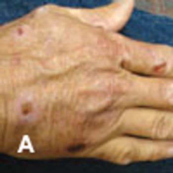
For several months, a 55-year-old white construction worker experienced intense burning of the skin when exposed to direct sunlight. In addition, multiple fragile blisters appeared on the dorsa of his hands and arms; these rapidly developed into crusted, superficial erosions.
In Dr David Kaplan's Dermclinic case of a keratoacanthoma in a 63-year-old woman (CONSULTANT, April 15, 2007, page 473), the lesion is referred to as a "low-grade squamous cell carcinoma." However, keratoacanthomas, while previously considered to be a variant of squamous cell carcinoma, are actually benign.
A 39-year-old woman complained of excruciating pain that radiated from a chronic lesion on the left upper lip to the entire left side of the face. She had AIDS but was not receiving antiretroviral therapy.
Dermclinic: A Photo Quiz to Hone Dermatologic Skills
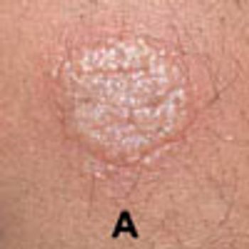
Tuberculin-type hypersensitivity is characterized by marked spongiotic dermatitis with intraepidermal and subepidermal vesiculation and scattered eosinophils.
Advertisement
Advertisement
Trending on Patient Care Online
1
From Amyloid Clearance to Daytime Function: Why Sleep Quality Matters for Brain Health
2
2026 Dyslipidemia Guideline Expands Statin Eligibility to 21.5 Million More US Adults
3
ACOG Releases New Guidance on HIV Screening and Prevention
4
Compulsive Smartphone Use Linked to Depressive Symptoms in Older Adults
5