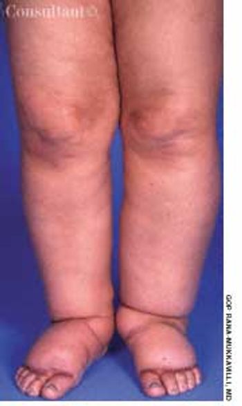
















































































































































During a routine office visit, a 64-year-old woman who has had type 2 diabetesfor more than 10 years complains of increased pedal edema. The edema is minimalon awakening and worsens throughout the day.


During a routine office visit, a 64-year-old woman who has had type 2 diabetesfor more than 10 years complains of increased pedal edema. The edema is minimalon awakening and worsens throughout the day.

How does amyloid in the pancreatic islets contribute tothe development and progression of type 2 diabetes?

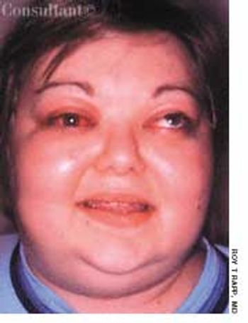
For 1 month, a 25-year-old woman had experienced discomfort in and around the left eye and diplopia. She was in good health; she reported no weight loss, excessive nervousness, heat intolerance, decreased strength, changes in the texture of hair or skin, or altered bowel habits. There was no personal or family history of goiter or other thyroid disease.

As the world of sport has embraced the participation of women and girls, the incidence of health problems that pertain specifically to premenopausal female athletes has increased significantly. One of these is the female athlete triad, which consists of 3 interrelated medical conditions associated with athletic training

Drs Sonia Arunabh and K. Rauhilla’s case of a 62-year-old woman with Raynaud’sphenomenon (CONSULTANT, September 15, 2001, page 1526) offers one ofthe finest photographs of this condition that I have seen (Figure).

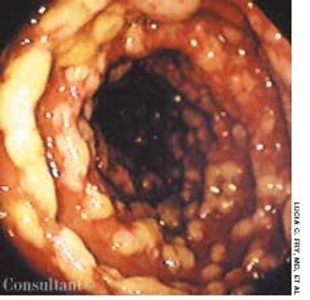
For 2 days, a 68-year-old woman had watery, yellowish diarrhea with mucus and left lower quadrant pain. Her medical history included hypertension, diabetes mellitus, and congestive heart failure (CHF); she had left the hospital 5 days earlier following treatment of an exacerbation of CHF with intravenous furosemide and sodium and fluid restriction. The patient was taking furosemide, lisinopril, and glipizide; she denied any recent antibiotic therapy.

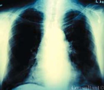
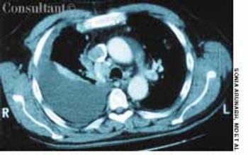
A 64-year-old woman with a history of diabetes, hypertension, and lymphoma was admitted to the hospital with a dull headache, conjunctival congestion, and slight dyspnea. Her pulse rate was 96 beats per minute; blood pressure, 146/68 mm Hg; and respiration rate, 22 breaths per minute. She also had increased jugular venous distention; cardiovascular and chest examination findings were normal. Edema of both arms and dilated blood vessels on the anterior chest wall were noted.

Heart failure(HF), the mostcommon Medicarediagnosisrelatedgroup,has a significant and growingimpact on health careresources. The incidenceof HF has tripled during thelast decade. Almost 5 millionAmericans have HF, and anestimated 500,000 new casesare diagnosed yearly. Thelifetime risk of HF is about20%.1 Drug therapy has improvedconsiderably in recentyears, but the magnitudeand severity of theproblem has created a needfor newer therapies--particularlysince HF is associatedwith an increased risk ofsudden death and a diminishedquality of life.2

Some degree of hyperpigmentationdevelops in most pregnantwomen. This coloration is more pronouncedin dark-skinned women; onnaturally pigmented areas, such as theareolae, perineum, and umbilicus; andon the axillae, inner thighs, and otherregions that are prone to friction.

Many of my older women patients think theyare at much higher risk for breast cancer thancoronary heart disease (CHD). How can we raise women'sawareness about their risks of CHD and therebyencourage them to take measures to prevent it?

Dr Gregory Rutecki's interactive teaching case, “A Middle-Aged Man WithPolyuria: The Initial Visit” (CONSULTANT, March 2001, page 357), provided awelcome opportunity for me to review the care I provide to my patients with type 2diabetes, who comprise a very large percentage of my practice.

Q:Should I avoid angiotensin-converting enzyme (ACE) inhibitors in mypatients with progressive renal insufficiency?

Q:Many of my patients appear to have white-coathypertension: their pressure is elevated whenmeasured in my office-but normal when measured athome. Am I ignoring significant hypertension if I do nottreat these patients? Or am I overtreating if I do treat?

Highly pruritic, 2- to 4-mm, papular lesions with central ulceration erupted on the back of a 66-year-old woman. She had had 2 similar outbreaks in the past. The patient was taking conjugated estrogens, alprazolam, and alendronate.

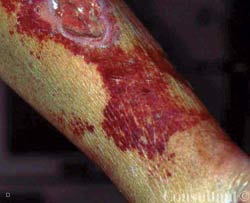
Cutaneous manifestations develop inapproximately 30% of persons withdiabetes. Premature atherosclerosisis a common complication of thedisease and can cause peripheral infarction,ulceration, and necrosis.

Affected infants present shortly afterbirth with a large bowel obstructionsecondary to transient dysmotility inthe descending colon. Although thecause is unknown, immaturity of thecolonic myenteric plexuses has beendemonstrated in some cases. Morethan 50% of affected infants are bornto mothers with diabetes. Other predisposingfactors include hypoglycemiaand sepsis.

This patient with long-standing insulin-dependent diabetes has difficultyclosing his hand because of “tight skin.” Diabetes is the cause: this findingoccurs more often in persons with microvascular complications, such asretinopathy and nephropathy. The condition may occur even in patients withwell-controlled diabetes.

Diabetic retinopathy is the most common cause of legal blindness in personsbetween the ages of 20 and 65 years. In this 56-year-old man with a 20-yearhistory of type 2 diabetes, multiple, scattered intraretinal (dot-blot) hemorrhagesand superficial nerve fiber layer (splinter) hemorrhages can be seen.An occasional Roth spot-an intraretinal hemorrhage with a white center thatrepresents a fibrin thrombus which occludes a ruptured blood vessel-is alsoevident. Numerous yellow, waxy, hard exudates are seen between the innerplexiform and inner nuclear layers of the retina. Cotton-wool spots are alsopresent, although no neovascularization is present.

This huge blister developed spontaneously on the foot of a woman withdiabetes mellitus. There was no area of redness around the blister, whichnormally occurs with burns and inflamed lesions. Spontaneous blisters andother skin manifestations are common in persons with diabetes.

Numerous brown spots on his leg brought this man with long-standing diabetes mellitus to his physician. He thought the spots were related to trauma.

This patient with long-standing insulin-dependent diabetes mellitus has difficulty in closing his hand because the skin is "very tight."

Numerous brown spots on the leg of a man with longstandingdiabetes are diabetic dermopathy. This is amicroangiopathy of the small arterioles of the legs, whichresults in atrophic brown papules.

The atrophic patches on the lower legs of this 47-year-old woman who has haddiabetes mellitus for over 20 years are those of necrobiosis lipoidica diabeticorum.These areas involve degeneration of collagen and elastic fibers in thelower dermis and changes in blood vessel walls. The lesions usually begin assingle or multiple elevated reddish nodules, most commonly in the pretibialarea. Over time, they expand and coalesce into distinctive brownish yellowpatches. These areas may be somewhat tender, but as they spread, theyfrequently become painless-unless they ulcerate. Because the involved skinis fragile, ulcers can form after any minor trauma.

Migraine is an episodic, often debilitatingcondition that affects women moreoften than men. Twenty-eight millionAmericans suffer from migraineheadaches-and nearly 75% of theseare women.1 Unlike other chronic painconditions, migraine has its peakprevalence during the years of greatestproductivity, when most women arejuggling family responsibilities andcareers.2 Many women are particularlysusceptible to migraine attacks justbefore and during menses.

For 3 days, a 23-yearold woman had been bothered by pruritic vaginal and vulvar lesions. A cheesy white vaginal discharge was associated with the itching. She denied being sexually active.