
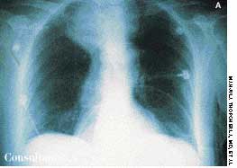
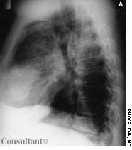
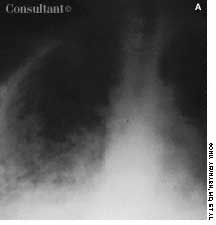
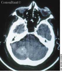

































































































































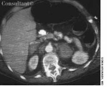
A 48-year-old woman was admitted to the hospital with deep venous thrombosis of the right leg. She had a history of non-Hodgkin lymphoma, which was in remission. The patient had been a heavy smoker for many years.
A 48-year-old woman was admitted to the hospital with deep venous thrombosis of the right leg. She had a history of non-Hodgkin lymphoma, which was in remission. The patient had been a heavy smoker for many years.
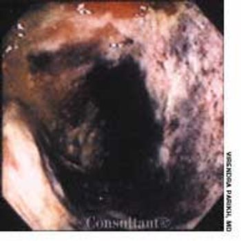
For 2 days, a 79-year-old man with a history of congestive heart failure experienced abdominal pain, diarrhea, and dark red rectal bleeding. The pain was localized to the left lower quadrant.
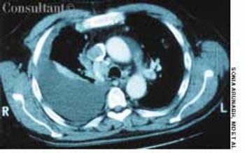
A 64-year-old woman with a history of diabetes, hypertension, and lymphoma was admitted to the hospital with a dull headache, conjunctival congestion, and slight dyspnea. Her pulse rate was 96 beats per minute; blood pressure, 146/68 mm Hg; and respiration rate, 22 breaths per minute. She also had increased jugular venous distention; cardiovascular and chest examination findings were normal. Edema of both arms and dilated blood vessels on the anterior chest wall were noted.
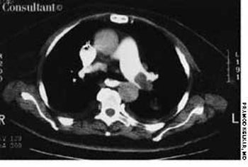
A 72-year-old woman had complained of shortness of breath for the past week. A CT scan showed a large filling defect in the left main pulmonary artery (Figure A, thin arrows) that extended into both the upper and lower branches. Another filling defect (not shown here) was seen along the posterior wall of the right main pulmonary artery, extending into the lower branch. These defects are compatible with pulmonary emboli.
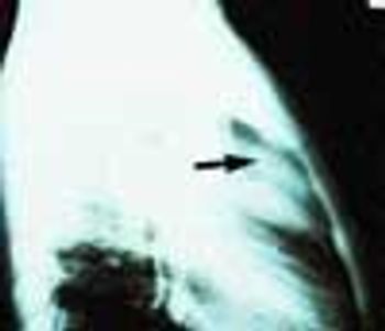
A 72-year-old woman who had fallen and injured the left side of her chest came to the emergency department complaining of pain in that area. She was physically stable and not short of breath. A soft systolic murmur was heard over the left precordium; the lungs were clear. A posteroanterior chest film showed no rib fracture but it did show an enlarged heart and a large, calcified ventricular aneurysm.
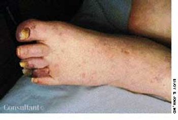
A 65-year-old man presented with bluish toes and petechiae on his toes and feet 3 days after having undergone renal angiography. Bilateral pitting pedal edema was noted, and laboratory tests revealed proteinuria, eosinophilia, and an erythrocyte sedimentation rate (ESR) of 65 mm/h.
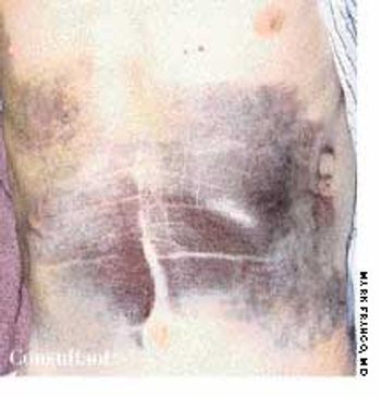
An 80-year-old man with a history of congestive heart failure, coronary artery disease, cardiomyopathy, and thoracic and abdominal aneurysms was taken to the emergency department because of mental status changes, back pain, and ecchymotic areas over his body. The ecchymoses started on his back 5 days before admission and spread to his abdomen.
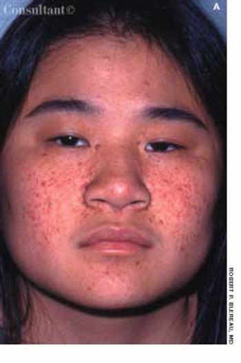
Tan-pink acneiform lesions on the face of a 15-year-old girl had not responded to topical acne therapy. A 1 × 0.5-cm, elevated subcutaneous nodule was noted on the right lateral knee. The lesions on her face and knee had been present for 11 years. The family history was noncontributory.
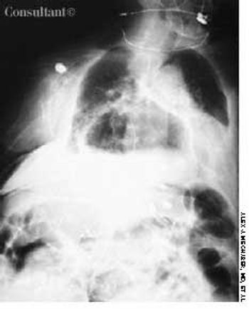
A 97-year-old woman with a history of hypertension and a paraesophageal hiatal hernia presented with abdominal distention and shortness of breath. Three days earlier, she had fallen and sustained a hairline pelvic fracture; she was evaluated in the emergency department and given narcotics for the pain. Subsequently, the patient's abdomen became increasingly distended, and she had no bowel movement for 3 days.
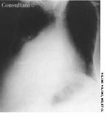
An 82-year-old woman was admitted to the hospital with severe dilated cardiomyopathy. The condition was alcohol-induced and had been diagnosed about 12 years earlier. The patient was taking angiotensin-converting enzyme inhibitors and diuretics.
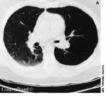
With a 1-year history of episodic wheezing, a 62-year-old woman (a smoker for the past 30 years) was being treated for bronchial asthma, but bronchodilator therapy did not control her symptoms. She was hospitalized with worsening dyspnea and a 4.5-kg (10-lb) weight loss over the past 3 months. There was no hemoptysis.
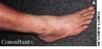
These orange-to-brown macules with red puncta, or cayenne pepper spots, are typical of Schamberg's disease (progressive pigmented purpuric dermatosis). The cause of this disorder is unknown, but it may be related to a cellular immune reaction or drug reaction.
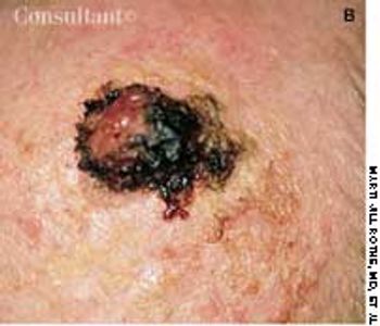
An 82-year-old man with Alzheimer's disease and atrial fibrillation was referred for evaluation of a lesion present for an undetermined period. It was initially noted 1 week earlier, at his first office visit with a geriatrician.

Cirrhosis and ascites developed in a 52-year-old man with a history of chronic hepatitis C and ethanol abuse. He was hospitalized because of bleeding esophageal varices, which were successfully treated with elastic band ligation.
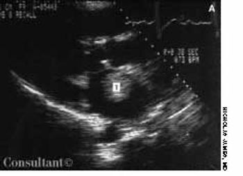
A 72-year-old man with a history of rheumatic heart disease presented with pulmonary congestion and syncope. Results of the cardiac examination suggested mitral stenosis and atrial fibrillation with a controlled ventricular response. Two-dimensional echocardi-ography revealed a large, free-floating ball thrombus (T) in the dilated left atrium and demonstrated rheumatic and severely stenotic mitral valve leaflets.

These collections of dilated vessels deep in the dermis and subcutaneous tissue are present at birth. They usually present as bluish or reddish lesions that are cystic, firm, and compressible. About 60% to 80% of cavernous hemangiomas undergo spontaneous involution, often with central clearing and fibrosis.

During a routine physical examination of a healthy 7-year-old African-American boy, this striking-and puzzling-hyperpigmentation was noted on his palms.
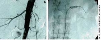
A 73-year-old man who had a history of long-standing essential hypertension, congestive heart failure, mild renal insufficiency, atrial fibrillation, and a mitral valve replacement presented with refractory hypertension. His medications included labetalol, irbesartan, and furosemide.
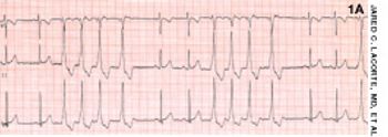
An 8-year-old boy was brought to his pediatrician for well-child care. On physical examination, an irregularly irregular heart rate was detected.
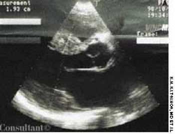
Dyspnea, orthopnea, and weight loss sent a 40-year-old woman for medical consultation. Fifteen years earlier, the patient had been nephrectomized because of left kidney lithiasis. There was no history of other symptoms or diseases.
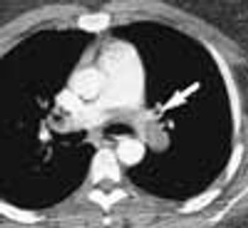
For 2 months, a 31-year-old woman had had dyspnea and dull, continuous retrosternal pain. She was admitted to the hospital, and a helical CT scan of the thorax identified a saddle pulmonary embolism. An ultrasonogram revealed deep venous thrombosis (DVT) in the left leg. Intravenous heparin was given; the patient was discharged, and warfarin was prescribed.
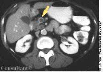
A 65-year-old woman with metastatic adenocarcinoma of the colon was undergoing chemotherapy following a colectomy and a hepatic wedge resection. The physical examination and laboratory data were unremarkable.
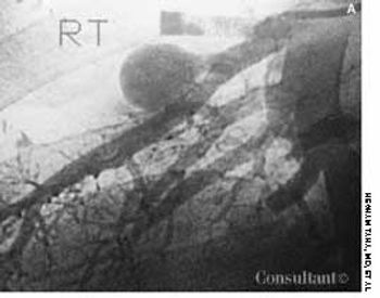
A 22-year-old man complained of progressive shortness of breath and abdominal distention. Three years before, he had completed chemotherapy for Hodgkin's disease and had since been in remission. Recently, he had been treated for tonsillitis with oral antibiotics.
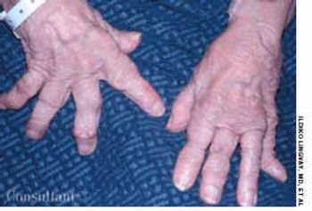
A 76-year-old woman had a 40-year history of rheumatoid arthritis (RA). She had repeatedly refused treatment with disease-modifying drugs, including methotrexate. Nodules began to develop 15 years after the initial diagnosis; they recurred after surgical removal.
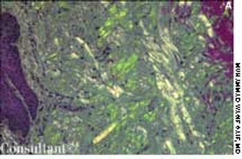
A 63-year-old African American man presented with orthopnea and pedal edema that had worsened during the past 4 months. Macroglossia was noted.